. 2011 Jul 1;10(13):2140-50.
doi: 10.4161/cc.10.13.16227. Epub 2011 Jul 1.

Scleroderma-like properties of skin from caveolin-1-deficient mice: implications for new treatment strategies in patients with fibrosis and systemic sclerosis

Affiliations

Scleroderma-like properties of skin from caveolin-1-deficient mice: implications for new treatment strategies in patients with fibrosis and systemic sclerosis

Remedios Castello-Cros et al. Cell Cycle. .

Abstract

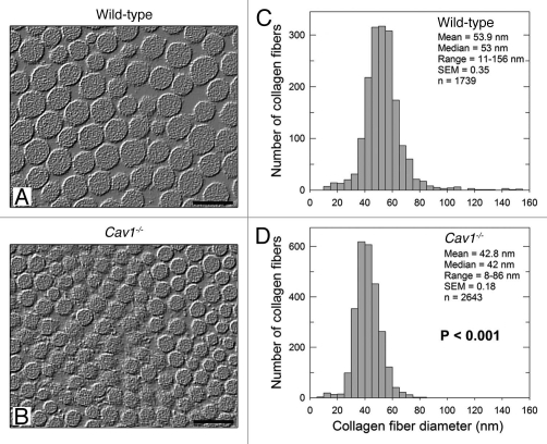
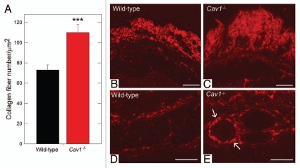
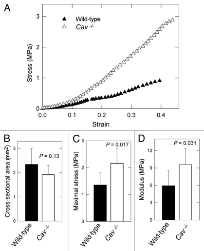
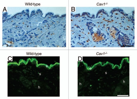
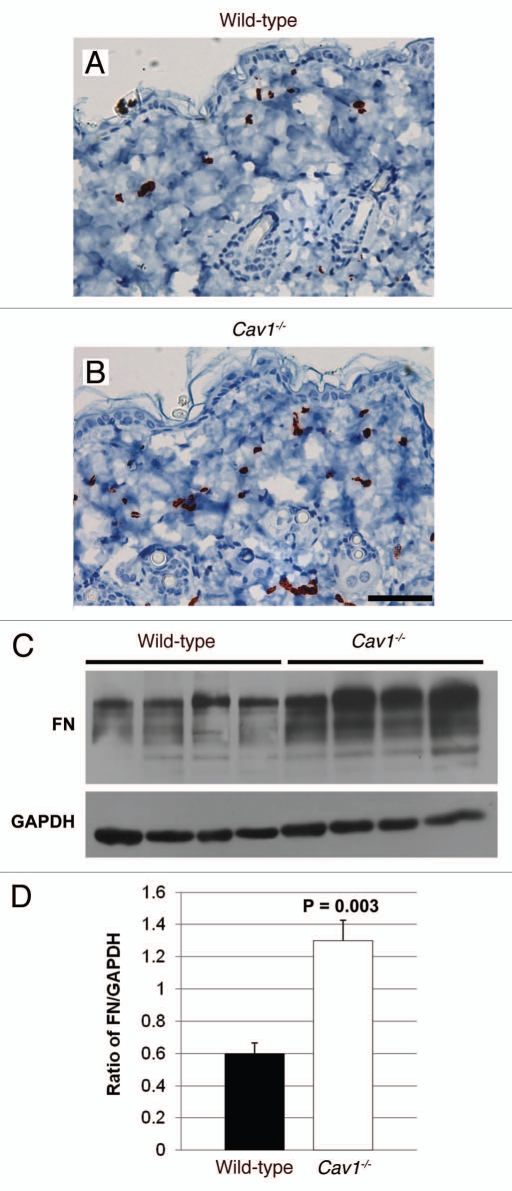
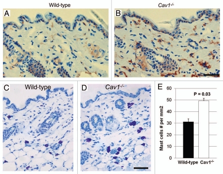
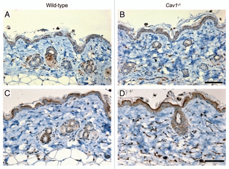
Caveolin-1 (Cav-1), the principal structural component of caveolae, participates in the pathogenesis of several fibrotic diseases, including systemic sclerosis (SSc). Interestingly, affected skin and lung samples from patients with SSc show reduced levels of Cav-1, as compared to normal skin. In addition, restoration of Cav-1 function in skin fibroblasts from SSc patients reversed their pro-fibrotic phenotype. Here, we further investigated whether Cav-1 mice are a useful pre-clinical model for studying the pathogenesis of SSc. For this purpose, we performed quantitative transmission electron microscopy, as well as biochemical and immuno-histochemical analysis, of the skin from Cav-1 (-/-) null mice. Using these complementary approaches, we now show that skin from Cav-1 null mice exhibits many of the same characteristics as SSc skin from patients, including a decrease in collagen fiber diameter, increased tensile strength, and stiffness, as well as mononuclear cell infiltration. Furthermore, an increase in autophagy/mitophagy was observed in the stromal cells of the dermis from Cav-1 (-/-) mice. These findings suggest that changes in cellular energy metabolism (e.g., a shift towards aerobic glycolysis) in these stromal cells may be a survival mechanism in this "hostile" or pro-inflammatory microenvironment. Taken together, our results demonstrate that Cav-1 (-/-) null mice are a valuable new pre-clinical model for studying scleroderma. Most importantly, our results suggest that inhibition of autophagy and/or aerobic glycolysis may represent a new promising therapeutic strategy for halting fibrosis in SSc patients. Finally, Cav-1 (-/-) null mice are also a pre-clinical model for a "lethal" tumor micro-environment, possibly explaining the link between fibrosis, tumor progression, and cancer metastasis.


Figures

Figure 1
Figure 1
Ultrastructural analysis of the skin from wild-type and Cav-1-/- mice. (A and B) Transmission electron microscopy (EM) micrographs of dermal collagen from wild-type (A) and Cav-1-/- (B) mice. (C and D) Distribution of diameters from dermal collagen from wild-type (C) and Cav-1-/- (D) mice. An Image J algorithm was designed to automatically obtain collagen fiber diameters from EM images (Sup. Fig. 1). Using this algorithm, the diameter of 1739 and 2643 collagen fibers was determined from wild-type and Cav-1-/- mice, respectively. Note that dermal collagen from Cav-1-/- mice exhibited a more compact and uniform pattern of fibril diameter and distribution, than wild-type. Indeed the mean and the median collagen diameters from Cav-1-/- mice were significantly smaller than those from wild-type mice (p < 0.001, as determined by Mann-Whitney Rank Sum Test using SigmaPlot). Scale bar = 100 nm.
Figure 2
Figure 2
Quantification of dermal collagen density in wild-type and Cav-1-/- mice. (A) Electron microscopy (EM) images of dermal collagen from wild-type and Cav-1-/- mice were analyzed with a custom Image J algorithm (Sup. Fig. 1). Using this approach, we determined the collagen density, as number of fibers per unit area. We observed that Cav-1-/- mice contain a significantly larger number of dermal collagen fibrils per unit area, than wild-type animals. (***p < 0.001, as determined by Mann-Whitney Rank Sum Test using SigmaPlot). (B–E) Picrosirius red staining of skin from wild-type and Cav-1-/- mice. Polarized light images of the dermis and the deeper dermis around skin appendages and blood vessels of wild-type (B and D) and Cav-1-/- mice (C and E) were stained with picrosirius red. Mature collagen fibers stained with picrosirius dye appear red when observed under polarized light, whereas less mature fibers, with fewer cross-links, appear yellow and green. Note that although both groups display mature collagen fibers in the dermis, a larger amount of collagen accumulation was found in the dermis, the deeper dermis, and around blood vessels of Cav-1-/- skin, as compared to wild-type mice. Scale bar = 200 µm.
Figure 3
Figure 3
Bio-mechanical properties of the skin from wild-type and Cav-1-/- mice. (A) Typical stress-strain response for wild-type versus Cav-1-/- dorsal skin. (B) No differences were found in the cross-sectional area between the two groups. (C and D) Determination of tensile strength and modulus in the skin in wild-type and Cav-1-/- mice. The maximum stress or “tensile strength” is the maximum amount of tensile stress that a body can be subjected to before failure and Young's modulus (slope of elastic region) is related to “stiffness,” the resistance of the material to elastic percent deformation caused by an applied stress. Skin from Cav-1-/- mice exhibited increased maximum stress (C) and modulus (D), than those from wild-type (C and D). Five animals of each genotype were analyzed. Data are reported as the mean ± SEM (p values are indicated in the graphs, as determined by the Student's t-test).
Figure 4
Figure 4
Cav-1-/- mice exhibit a net change in collagen synthesis. (A and B) Prolyl-4-hydroxylase (P4HB) expression in the skin from wild-type (A) and Cav-1-/- (B) mice. P4HB is a key enzyme in collagen biosynthesis that catalyzes the 4-hydroxylation of prolyl residues. Immuno-staining indicates positive labeling of the cells expressing P4HB. Representative images are shown from three animals per group. Note that the dermis of Cav-1-/- mice contains an increased number of P4HB positive cells. In addition, the P4HB positive cells in Cav-1-/- mice exhibited a marked increased expression of this enzyme. (C and D) In situ collagenase activity was determined using DQ-collagen type I, as substrate in skin from wild-type (B) and Cav-1-/- mice (C). Multiple cryostat sections of the skin from these animals were incubated overnight with DQ-Collagen type I, dissolved in LGT-agarose. Fluorescence due to collagenase activity (green) was found in the epidermis (e), stromal cells of the dermis (s) and in hair follicles (h). Note that we observed similar collagenase activity in the skin from both wild-type and Cav-1-/- mice. Take together, this data suggest that the skin from Cav-1-/- mice may displays a net increase in collagen synthesis and/or accumulation. Scale bar = 50 microns.
Figure 5
Figure 5
Immunohistochemical detection of myo-fibroblasts in the skin of wild-type and Cav-1-/- mice. Skin sections from wild-type (A) and Cav-1-/- (B) mice were stained with α-SMA, a myo-fibroblast cell marker. Immuno-staining indicates positive labeling of myo-fibroblasts. Representative images are shown from three animals per group. Note that an increased number of α-SMA positive cells was observed in the dermis of Cav-1-/- mice, as compared to normal wild-type skin. (C) Analysis of fibronectin expression in the skin of wild-type and Cav-1-/- mice. Western blots of total protein extracted from wild-type and Cav-1-/- mice shows the expression of fibronectin (FN) and the loading control glyceraldehydes-3-phosphate dehydrogenase (GAPDH). (D) Quantification of fibronectin expression in the in the skin of wild-type and Cav-1-/- mice. Intensity of the western blot bands for fibronectin and GAPDH were quantified using NIH Image J. Note that a significant increased in fibronectin expression was observed in the Cav-1-/- skin, as compared to normal wild-type mouse skin. Results are indicated as the mean ± SEM. p values are indicated in the graphs, as determined by the Student's t-test. Scale bar = 50 microns.
Figure 6
Figure 6
Inflammatory cell content of the skin in wild-type and Cav-1-/- mice. (A and B) Frozen sections from the skin were stained with the F4/80 antibody, a macrophage cell marker, to evaluate macrophage infiltration in the dermis of wild-type (A) and Cav-1-/- mice (B). Immuno-staining indicates positive labeling of macrophages. (C and D) Histochemical staining to detect mast cells. Using this approach mast cells stain purple. Scale bar = 50 microns. (E) Quantification of mast cells in the skin from wild-type and Cav-1-/- mice. The number of mast cells was determined in five equal areas of skin (n = 5), from the sub-endothelium to the muscle. Note that increased infiltration of macrophages and mast cells was observed in the dermis of Cav-1-/- mice, as compared with normal wild-type dermis. Results are indicated as the mean ± SEM. p values are indicated in the graphs, as determined by the Student's t-test.
Figure 7
Figure 7
Autophagy/mitophagy in stromal cells from the dermis of wild-type and Cav-1-/- mice. Paraffin-embedded sections from the skin were stained with a LC3 antibody, to evaluate the degree of autophagy in stromal cells in the dermis of wild-type (A) and Cav-1-/- mice (B). Note that an increased number of stromal cells are positive for LC3 in the dermis of Cav-1-/- mice, in comparison to wild-type mice. The skin sections of these two groups were also stained with BNIP3L, a mitophagy marker. A marked increased in the number of dermal stromal cells that are positive for BNIP3L was observed in Cav-1-/- mice (D), in comparison with wild-type mice (C).

Comment in

References

    1. Jimenez SA, Derk CT. Following the molecular pathways toward an understanding of the pathogenesis of systemic sclerosis. Ann Intern Med. 2004;140:37–50. - PubMed
    1. LeRoy EC. Increased collagen synthesis by scleroderma skin fibroblasts in vitro: A possible defect in the regulation or activation of the scleroderma fibroblast. J Clin Invest. 1974;54:880–889. - PMC - PubMed
    1. Jimenez SA, Feldman G, Bashey RI, Bienkowski R, Rosenbloom J. Co-ordinate increase in the expression of type I and type III collagen genes in progressive systemic sclerosis fibroblasts. Biochem J. 1986;237:837–843. - PMC - PubMed
    1. Jimenez SA, Bashey RI. Collagen synthesis by scleroderma fibroblasts in culture. Arthritis Rheum. 1977;20:902–903. - PubMed
    1. Takeda K, Hatamochi A, Ueki H, Nakata M, Oishi Y. Decreased collagenase expression in cultured systemic sclerosis fibroblasts. J Invest Dermatol. 1994;103:359–363. - PubMed

Publication types

MeSH terms