. 2012 Jan;69(1):165-73.
doi: 10.1007/s00018-011-0744-0. Epub 2011 Jun 14.

Mechanisms regulating cilia growth and cilia function in endothelial cells

Affiliations

Mechanisms regulating cilia growth and cilia function in endothelial cells

Shakila Abdul-Majeed et al. Cell Mol Life Sci. 2012 Jan.

Abstract

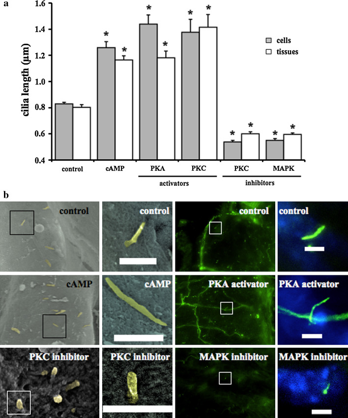
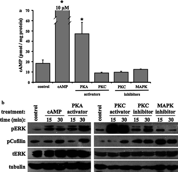
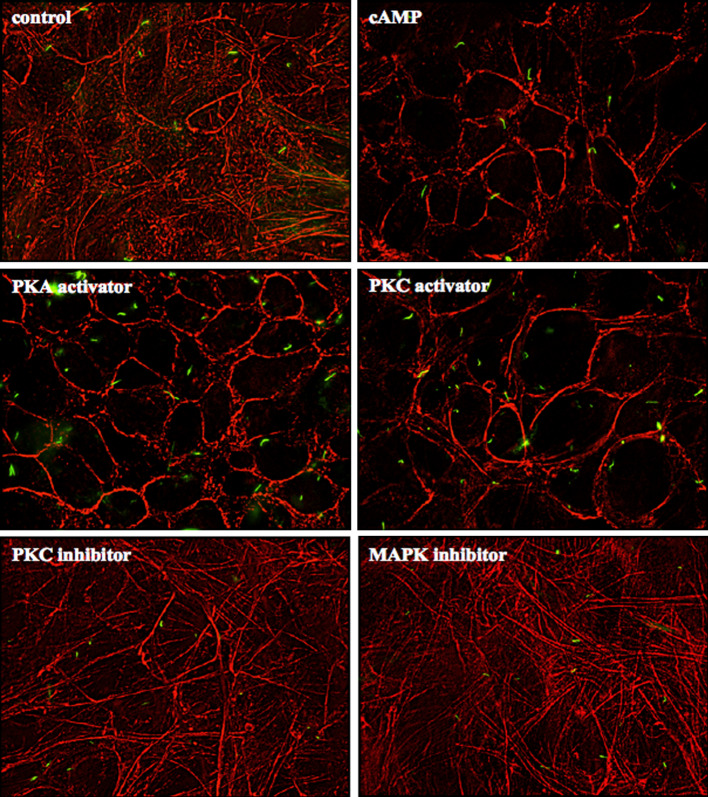
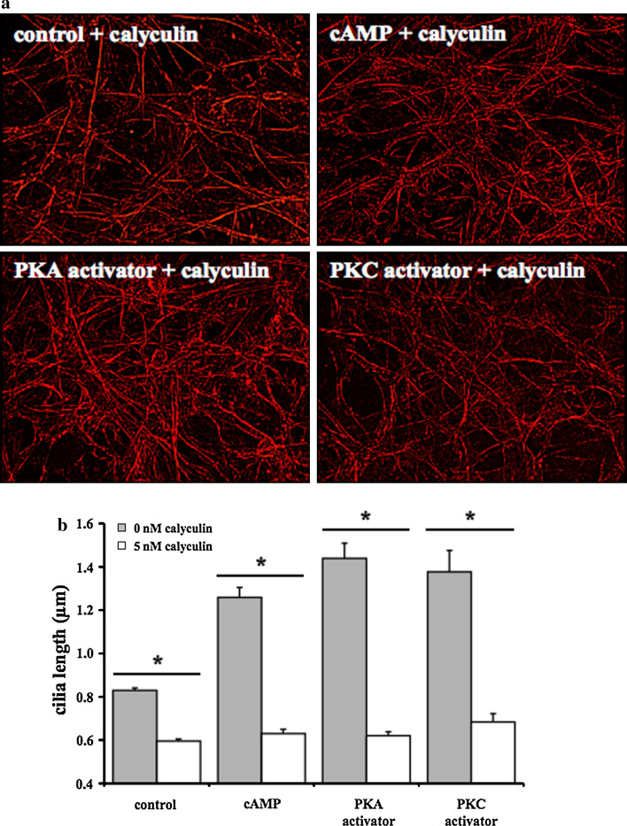
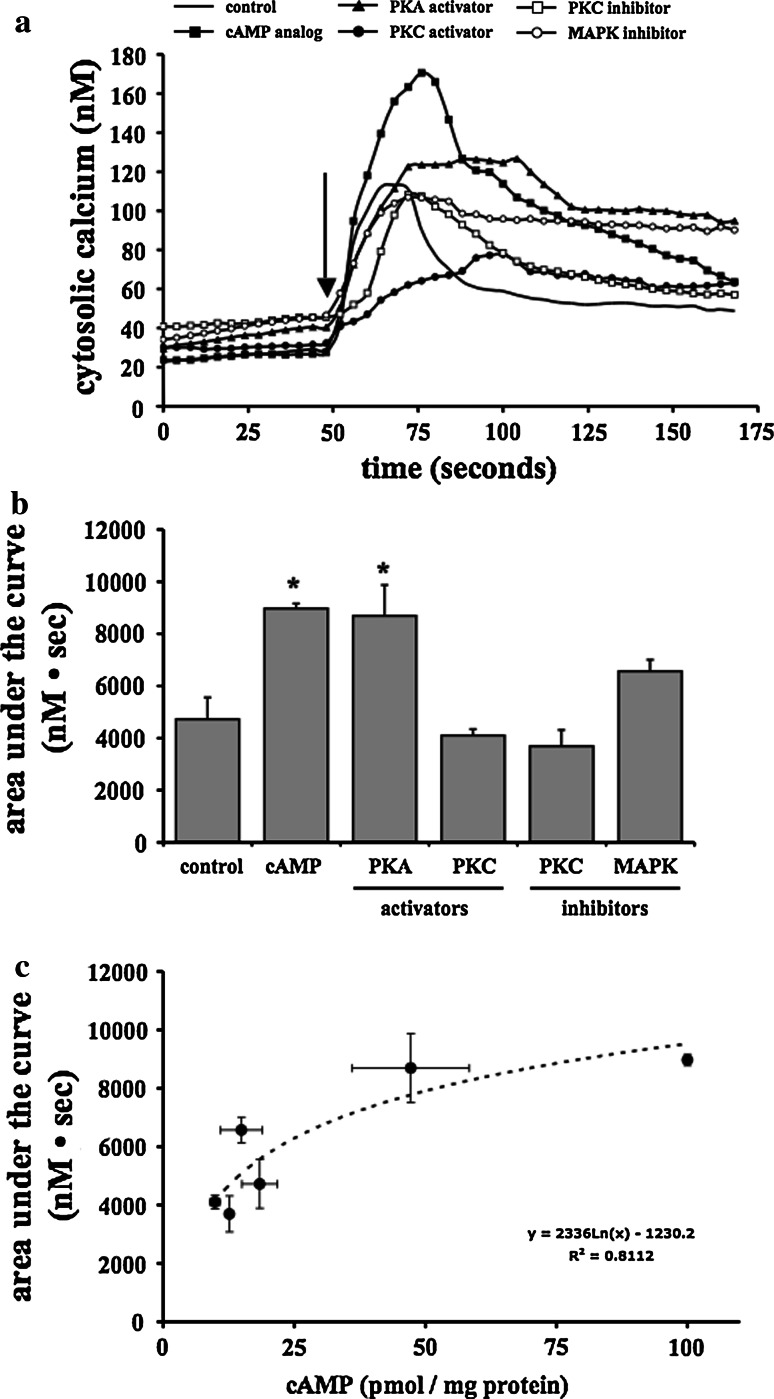
The primary cilium is an important sensory organelle present in most mammalian cells. Our current studies aim at examining intracellular molecules that regulate cilia length and/or cilia function in vitro and ex vivo. For the first time, we show that intracellular cAMP and cAMP-dependent protein kinase (PKA) regulate both cilia length and function in vascular endothelial cells. Although calcium-dependent protein kinase modulates cilia length, it does not play a significant role in cilia function. Cilia length regulation also involves mitogen-activated protein kinase (MAPK), protein phosphatase-1 (PP-1), and cofilin. Furthermore, cofilin regulates cilia length through actin rearrangement. Overall, our study suggests that the molecular interactions between cilia function and length can be independent of one another. Although PKA regulates both cilia length and function, changes in cilia length by MAPK, PP-1, or cofilin do not have a direct correlation to changes in cilia function. We propose that cilia length and function are regulated by distinct, yet complex intertwined signaling pathways.


Figures

Fig. 1
Fig. 1
Cilia length can be modulated with pharmacological agents. a Length of primary cilia in mono-layered cells and femoral arteries was measured before (control) and after treatment with 8-pCPT-cAMP (cAMP, 10 μmol/l), forskolin (PKA activator, 10 μmol/l), PMA (PKC activator, 0.5 μmol/l), bisindolylmaleimide (PKC inhibitor, 0.5 μmol/l), or PD98059 (MAPK inhibitor, 10 μmol/l) for 16 h. The cAMP analog and activators significantly increased cilia length, while the inhibitors significantly decreased cilia length. b Representative scanning electron and immunofluorescence images from femoral arteries are shown. Acetylated-α-tubulin was used to identify the primary cilia. Boxes indicates a greater magnification from the field of view. Bar 1 μm. n = 3 independent experiments; over 120 cilia were measured in cultured cells, and about 50 cilia were measured in femoral arteries. *p < 0.05 compared to control group
Fig. 2
Fig. 2
Ciliary length modulation involves a combination of cAMP, pERK, and cofilin activation. a Intracellular cAMP was measured in untreated cells (control) and cells treated with 8-pCPT-cAMP (cAMP), forskolin (PKA activator), PMA (PKC activator), bisindolylmaleimide (PKC inhibitor), or PD98059 (MAPK inhibitor). b Western blots depicting phosphorylated ERK (pERK), phospho-cofilin (pCofilin), total ERK (tERK), and α-tubulin (tubulin) are shown in cells with corresponding treatments for 15 or 30 min. α-tubulin was used as a loading control. n = 3 independent experiments for cAMP measurement and two for Western blots. *p < 0.05 compared to control group
Fig. 3
Fig. 3
Ciliary length modulation coincides with actin rearrangement. Representative images of primary cilia (green) and cytoskeletal actin filament (red) are shown. Actin filament was examined with phalloidin in untreated cells (control) and cells treated with 8-pCPT-cAMP (cAMP), forskolin (PKA activator), PMA (PKC activator), bisindolylmaleimide (PKC inhibitor), or PD98059 (MAPK inhibitor). Filamentous actin stress fibers are reorganized to form cortical actin in cells treated with cAMP, PKA activator, or PKC activator. On the other hand, cells treated with PKC inhibitor or MAPK inhibitor did not show apparent difference in actin stress fibers when compared to control. n = 3 independent experiments
Fig. 4
Fig. 4
Inhibition of actin rearrangement is sufficient to shorten ciliary length. a Cytoskeletal actin filament was analyzed in cells before and after treatment with calyculin, a protein phosphatase-1 inhibitor. Regardless of the absence or presence of cAMP analog, PKA activator, or PKC activator, filamentous actin stress fibers are always observed in cells treated with calyculin. b Calyculin-induced actin stress fiber maintenance is accompanied by a shortening in cilia length. n = 3 independent experiments. *p < 0.05
Fig. 5
Fig. 5
Cilia function is regulated by cAMP-PKA activity. a Changes in cytosolic calcium in response to fluid-flow shear stress were determined in untreated cells (control) and cells treated with 8-pCPT-cAMP (cAMP analog), forskolin (PKA activator), PMA (PKC activator), bisindolylmaleimide (PKC inhibitor), or PD98059 (MAPK inhibitor). Arrow indicates the start of fluid shear stress. b Cilia function quantified through total changes in cytosolic calcium is indicated as area under the curve (nM s). Activation of cAMP-dependent protein kinase considerably promotes cilia function. c Intracellular cAMP concentrations (pmol/mg protein) shows a functional correlation with cilia function, indicated as area under the curve (nM s). n ≥ 3 independent experiments; each represents an average of 100–150 cells
Fig. 6
Fig. 6
Cilia length and function are hypothesized to a complex divergence cellular pathway. Our working model predicts that both calcium- and cAMP-dependent protein kinases (PKC and PKA) are involved in regulating cilia length through MAP kinase (MAPK) and protein phosphatase-1 (PP-1). Our data further indicates that PP-1 plays an important role in actin rearrangement, which is a requirement for cilia length regulation. In addition to its role in cilia length modulation, PKA is also involved in regulating cilia function. Because PKC and MAPK are not involved in regulating cilia function, we propose that cilia length and function can be regulated by complex intertwined signaling pathways

References

    1. Singla V, Reiter JF. The primary cilium as the cell’s antenna: signaling at a sensory organelle. Science. 2006;313(5787):629–633. doi: 10.1126/science.1124534. - DOI - PubMed
    1. Nauli SM, Haymour HS, AbouAlaiwi WA, Lo ST, Nauli AM (2011) Primary cilia are mechanosensory organelles in vestibular tissues. In: Mechanosensitivity and mechanotransduction, Chap 14. Springer, Berlin Heidelberg New York. ISBN: 978-90-481-9880-1
    1. Brown NE, Murcia NS. Delayed cystogenesis and increased ciliogenesis associated with the re-expression of polaris in Tg737 mutant mice. Kidney Int. 2003;63(4):1220–1229. doi: 10.1046/j.1523-1755.2003.00863.x. - DOI - PubMed
    1. Pitaval A, Tseng Q, Bornens M, Thery M. Cell shape and contractility regulate ciliogenesis in cell cycle-arrested cells. J Cell Biol. 2010;191(2):303–312. doi: 10.1083/jcb.201004003. - DOI - PMC - PubMed
    1. Resnick A, Hopfer U. Force-response considerations in ciliary mechanosensation. Biophys J. 2007;93(4):1380–1390. doi: 10.1529/biophysj.107.105007. - DOI - PMC - PubMed

Publication types

MeSH terms

LinkOut - more resources