. 2011 Oct;22(10):1179-90.
doi: 10.1089/hum.2011.069. Epub 2011 Aug 10.

Long-term preservation of cones and improvement in visual function following gene therapy in a mouse model of leber congenital amaurosis caused by guanylate cyclase-1 deficiency

Affiliations

Long-term preservation of cones and improvement in visual function following gene therapy in a mouse model of leber congenital amaurosis caused by guanylate cyclase-1 deficiency

Marija Mihelec et al. Hum Gene Ther. 2011 Oct.

Abstract

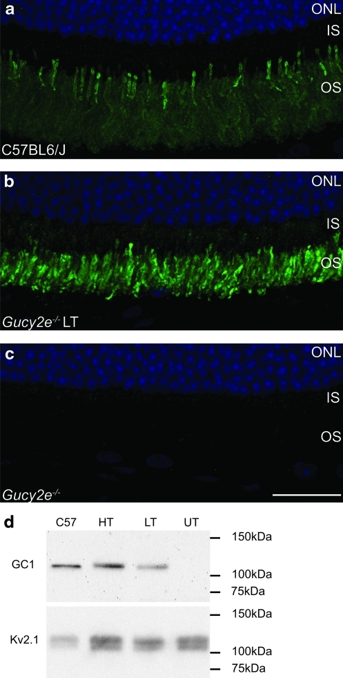
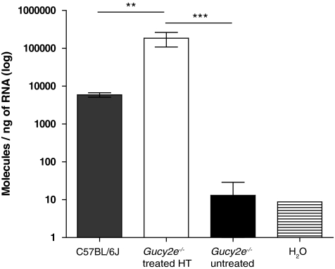
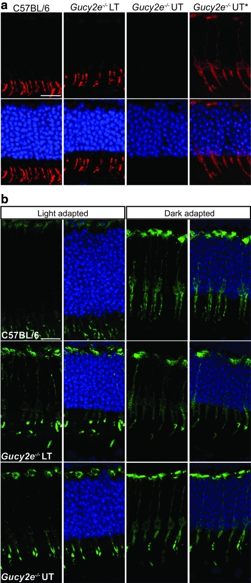
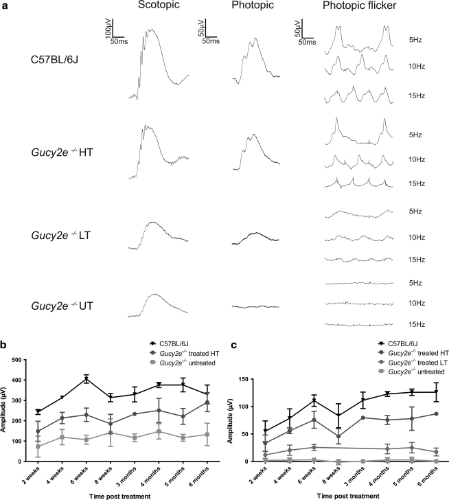
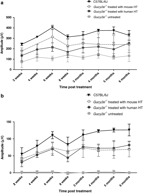
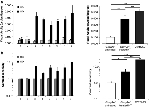
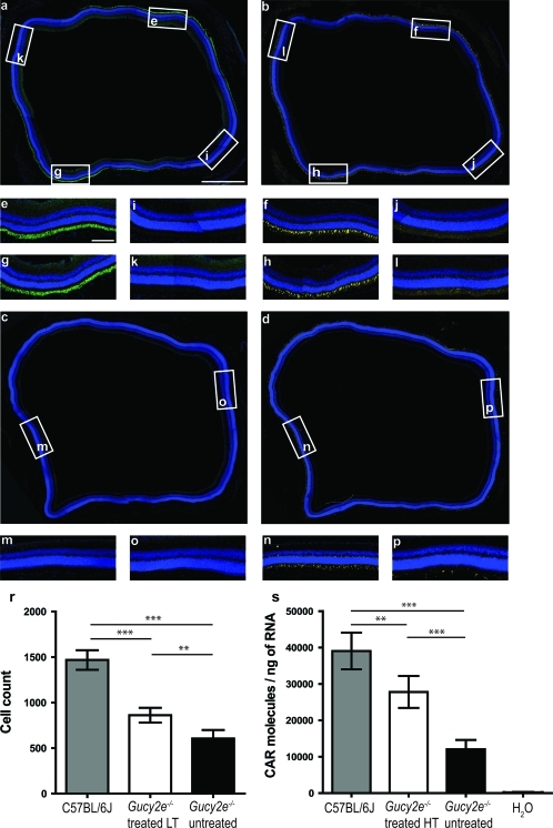
Leber congenital amaurosis (LCA) is a severe retinal dystrophy manifesting from early infancy as poor vision or blindness. Loss-of-function mutations in GUCY2D cause LCA1 and are one of the most common causes of LCA, accounting for 20% of all cases. Human GUCY2D and mouse Gucy2e genes encode guanylate cyclase-1 (GC1), which is responsible for restoring the dark state in photoreceptors after light exposure. The Gucy2e(-/-) mouse shows partially diminished rod function, but an absence of cone function before degeneration. Although the cones appear morphologically normal, they exhibit mislocalization of proteins involved in phototransduction. In this study we tested the efficacy of an rAAV2/8 vector containing the human rhodopsin kinase promoter and the human GUCY2D gene. Following subretinal delivery of the vector in Gucy2e(-/-) mice, GC1 protein was detected in the rod and cone outer segments, and in transduced areas of retina cone transducin was appropriately localized to cone outer segments. Moreover, we observed a dose-dependent restoration of rod and cone function and an improvement in visual behavior of the treated mice. Most importantly, cone preservation was observed in transduced areas up to 6 months post injection. To date, this is the most effective rescue of the Gucy2e(-/-) mouse model of LCA and we propose that a vector, similar to the one used in this study, could be suitable for use in a clinical trial of gene therapy for LCA1.


Figures

FIG. 1.
FIG. 1.
Correct size and localization of guanylate cyclase-1 (GC1) protein in treated Gucy2e–/– eyes. GC1 immunofluorescence (green) was detected in the outer segments of photoreceptor cells in (a) wild-type eyes and (b) Gucy2e–/– eyes treated with low-titer (LT) vector. (c) No staining was observed in untreated Gucy2e–/– eyes. 4′,6-Diamidino-2-phenylindole (DAPI) nuclear counterstaining is shown in blue. (d) Western blot showing 120-kDa GC1 protein in Gucy2e–/– eyes treated with low-titer (LT) or high-titer (HT) vector and in C57BL/6J wild-type controls, but not in Gucy2e–/– untreated eyes (UT). Kv2.1 was used as a loading control. OS, outer segments; IS, inner segments; ONL, outer nuclear layer. Scale bar: 25 μm. Color images available online at www.liebertonline.com/hum
FIG. 2.
FIG. 2.
GUCY2D transcript levels in Gucy2e–/– eyes treated with high-titer vector. Real-time RT-PCR analyses comparing levels of GUCY2D transcript in high-titer vector-treated Gucy2e–/– eyes with endogenous Gucy2e transcript in C57BL/6J wild-type eyes and untreated Gucy2e–/– eyes. The levels of introduced GUCY2D transcript in treated eyes are 30-fold higher than those seen in C57BL/6J wild-type control eyes (p<0.01, one-way ANOVA). Transcript levels detected in untreated controls are significantly lower than the levels of GUCY2D transcript detected in treated Gucy2e–/– eyes (p<0.001, one-way ANOVA).
FIG. 3.
FIG. 3.
Cone α-transducin levels and localization are restored in Gucy2e–/– treated eyes. C57BL/6J wild-type, Gucy2e–/– eyes treated with low-titer (LT) vector and untreated (UT) retinas were stained with antibodies against cone α-transducin (a, red) and cone arrestin (b, green). (a) Restored cone α-transducin localization to the outer segments of cones in the treated eyes, comparable to wild-type eyes. The images in the first three columns were taken using the same microscope settings, whereas the images in the last column, designated as Gucy2e–/– UT*, are identical to those in the Gucy2e–/– UT column except that they were taken with a long exposure and lighting levels were manipulated in Adobe Photoshop 7.0 Elements to reveal the signal in the red channel. The results indicate substantially reduced levels of cone α-transducin in untreated Gucy2e–/– eyes, and restored levels in treated eyes. (b) The two left-hand columns are images of retinal sections of eyes harvested and processed under normal light conditions and the right-hand columns are images of those harvested and processed in the dark. In the light, cone arrestin is localized in the cone outer segments and synaptic regions of wild-type as well as treated and untreated Gucy2e–/– eyes. In the dark, cone arrestin is localized throughout the cone cells in wild-type eyes, and in treated and untreated Gucy2e–/– eyes. DAPI nuclear counterstain is shown in blue. Scale bars: 15 μm. Color images available online at www.liebertonline.com/hum
FIG. 4.
FIG. 4.
Improvement in cone and rod function in treated Gucy2e–/– eyes. (a) Representative electroretinographic (ERG) traces from C57BL/6J wild-type mice, Gucy2e–/– mice 6 months after treatment with high-titer (HT) or low-titer (LT) vector, and untreated Gucy2e–/– (UT) mice. Rod-mediated b-wave responses of Gucy2e–/– animals treated with low-titer vector were no different from those of untreated eyes, whereas responses of animals treated with high-titer vector were restored to an average of 65% of wild-type amplitudes. Cone-mediated ERG of Gucy2e–/– animals treated with low-titer vector improved on average to 20% of wild-type amplitudes, whereas the responses of animals treated with high-titer vector were restored to 65% compared with wild-type eyes. (b) Over time, scotopic b-wave amplitudes in Gucy2e–/– mice treated with high-titer vector were significantly higher than those in untreated mice (p=0.0005, two-way ANOVA). (c) Data from animals treated with low-titer vector were significantly different from those of untreated and wild-type eyes at all time points examined (p<0.05, two-way ANOVA). Cone-mediated ERG responses of eyes treated with high-titer vector were significantly higher than those of untreated eyes at all time points (p<0.001, two-way ANOVA) and significantly lower than in wild-type eyes at all time points (p<0.05, two-way ANOVA), except at 2 weeks post injection.
FIG. 5.
FIG. 5.
Comparison of photoreceptor function restoration after transfer of human GUCY2D and mouse Gucy2e transgenes. (a) Consistent rod ERG improvement was observed in eyes treated with both vectors (mean±SD). No statistically significant difference was observed between the efficacy of rescue using both vectors (p=0.587, two-way ANOVA). (b) Cone-mediated ERG b-wave responses of Gucy2e–/– eyes treated with the mouse transcript did not differ from those treated with the human transcript (p=0.9887, two-way ANOVA). Cone-mediated ERG responses achieved with the mouse transcript were approximately 62% of the response seen in C57BL/6J wild-type eyes, compared with 65% achieved with the human transgene.
FIG. 6.
FIG. 6.
Restoration of cone-mediated optomotor behavior in Gucy2e–/– treated eyes. Shown is the photopic visual acuity and contrast sensitivity measurements in two untreated Gucy2e–/– mice (animals 1 and 2), and six mice treated with high-titer vector (animals 3–8) 4 months post treatment. Results are presented for each animal and averaged per group in the right-hand graphs. (a) Robust improvement in visual acuity was consistently observed in treated eyes (p<0.0001, one-way ANOVA). (b) A significant improvement in contrast sensitivity was detected in treated eyes (p<0.05, one-way ANOVA). OD, right eye; OS, left eye.
FIG. 7.
FIG. 7.
Cone preservation in the transduced areas of Gucy2e–/– treated eyes. Shown are adjoining retinal sections of Gucy2e–/– eyes 6 months post treatment with low-titer vector (a and b) and untreated contralateral eyes (c and d) stained with antibodies against GC1 (a and c, green) and cone α-transducin (b and d, yellow). (el) Insets are images of corresponding regions outlined in (a) and (b). In the areas where GC1 staining is evident in the treated eyes (e and g) there is also expression of cone α-transducin (f and h). In the untransduced areas, where there is no GC1 (i and k), there is no cone α-transducin staining (j and l). (m and n) Images of the superior retina, and (o and p) of the inferior retina of the untreated eye (c and d). There is no GC1 staining in the untreated eye (c). Weak cone α-transducin staining is evident in the superior untreated retina (n) with few cone cells present in the inferior part of the retina (p). Scale bars: (a) 500 μm; (e) 100 μm. (r) Number of cone cells determined by cone α-transducin staining in Gucy2e–/– eyes treated with low-titer vector, compared with untreated Gucy2e–/– eyes and C57BL/6J wild-type controls. The cone number in treated eyes was 20% higher than in untreated eyes (p<0.001, one-way ANOVA). (s) Real-time RT-PCR quantification of cone arrestin mRNA (CAR) in wild-type, Gucy2e–/– untreated eyes and eyes 5 months post treatment with high-titer vector. The levels of cone arrestin are 1.3 times higher in treated eyes compared with untreated eyes (p<0.0001, one-way ANOVA) and have increased to 70% of the levels in wild-type eyes. Color images available online at www.liebertonline.com/hum

Comment in

References

    1. Acland G.M. Aguirre G.D. Ray J., et al. Gene therapy restores vision in a canine model of childhood blindness. Nat. Genet. 2001;28:92–95. - PubMed
    1. Alexander J.J. Umino Y. Everhart D., et al. Restoration of cone vision in a mouse model of achromatopsia. Nat. Med. 2007;13:685–687. - PMC - PubMed
    1. Ali R.R. Sarra G.M. Stephens C., et al. Restoration of photoreceptor ultrastructure and function in retinal degeneration slow mice by gene therapy. Nat. Genet. 2000;25:306–310. - PubMed
    1. Allocca M. Mussolino C. Garcia-Hoyos M., et al. Novel adeno-associated virus serotypes efficiently transduce murine photoreceptors. J. Virol. 2007;81:11372–11380. - PMC - PubMed
    1. Baehr W. Karan S. Maeda T., et al. The function of guanylate cyclase 1 and guanylate cyclase 2 in rod and cone photoreceptors. J. Biol. Chem. 2007;282:8837–8847. - PMC - PubMed

Publication types

MeSH terms