. 2011 Jul 29;286(30):26533-40.
doi: 10.1074/jbc.M111.265322. Epub 2011 Jun 14.

BCL-2 modifying factor (BMF) is a central regulator of anoikis in human intestinal epithelial cells

Affiliations

BCL-2 modifying factor (BMF) is a central regulator of anoikis in human intestinal epithelial cells

Martin Hausmann et al. J Biol Chem. .

Abstract

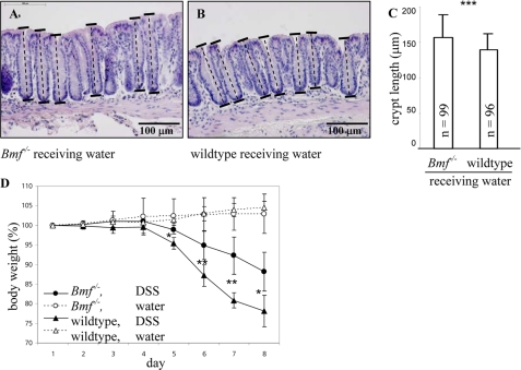
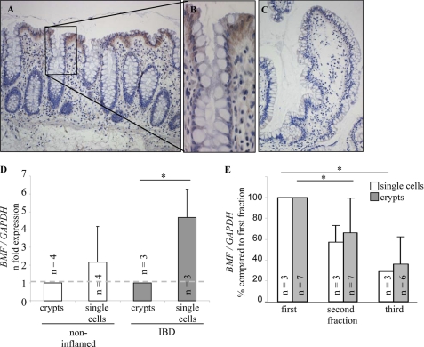
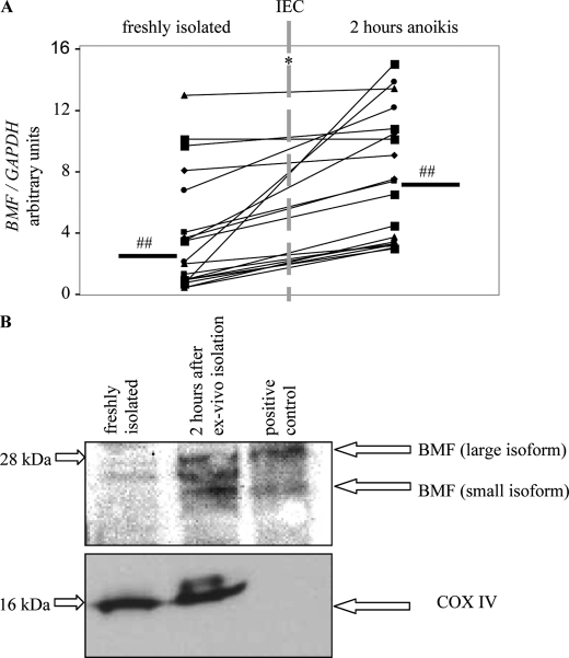
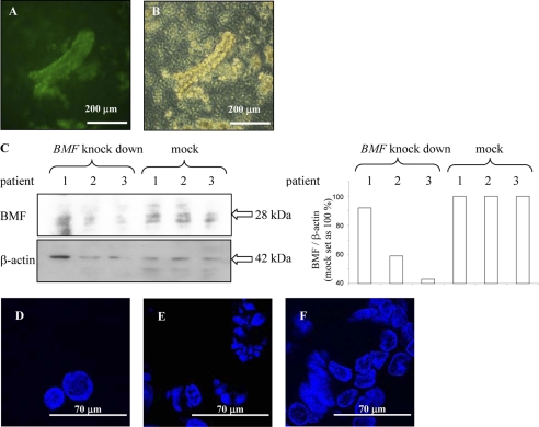
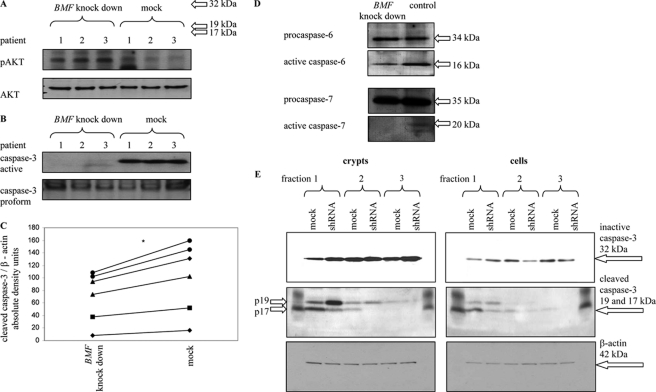
BCL-2 modifying factor (BMF) is a sentinel considered to register damage at the cytoskeleton and to convey a death signal to B-cell lymphoma 2. B-cell lymphoma 2 is neutralized by BMF and thereby facilitates cytochrome C release from mitochondria. We investigated the role of BMF for intestinal epithelial cell (IEC) homeostasis. Acute colitis was induced in Bmf-deficient mice (Bmf(-/-)) with dextran sulfate sodium. Colonic crypt length in Bmf(-/-) mice was significantly increased as compared with WT mice. Dextran sulfate sodium induced less signs of colitis in Bmf(-/-) mice, as weight loss was reduced compared with the WT. Primary human IEC exhibited increased BMF in the extrusion zone. Quantitative PCR showed a significant up-regulation of BMF expression after initiation of anoikis in primary human IEC. BMF was found on mitochondria during anoikis, as demonstrated by Western blot analysis. RNAi mediated knockdown of BMF reduced the number of apoptotic cells and led to reduced caspase 3 activity. A significant increase in phospho-AKT was determined after RNAi treatment. BMF knockdown supports survival of IEC. BMF is induced in human IEC by the loss of cell attachment and is likely to play an important role in the regulation of IEC survival.


Figures

FIGURE 1.
FIGURE 1.
Crypt length and body weight loss. Crypt lengths for Bmf−/− mice receiving water (A) and wild-type mice receiving water (B). A section 0–1 cm from the distal end of the colon is shown. C, a normality test for crypt length in Bmf−/− mice and wild-type mice receiving water failed, and a Mann-Whitney rank sum test was performed. Bmf−/− mice showed a significantly increased crypt length compared with wild-type mice. ***, p < 0.001. D, Bmf−/− mice (○ and ●) and wild-type mice (△ and ▴) received either DSS (▴) or water (△). Bars represent mean ± S.D. One-way analysis of variance was used. Body weight loss was significantly different for wild-type mice upon DSS treatment compared with Bmf−/− mice upon DSS treatment on days 5, 6, 7, and 8. Day 5, p = 0.016; day 6, p = 0.008; day 7, p = 0.007; and day 8, p = 0.016; n = 5 each. Bmf−/− mice showed a significantly decreased body weight loss upon DSS treatment and increased crypt length upon water treatment.
FIGURE 2.
FIGURE 2.
Immunohistochemistry for BMF on sections from human (A and B) large intestine and (C) small intestine. A and B, immunostaining of BMF on section from a Crohn's disease patient without inflammation. C, negative control without primary antibody. Immunohistochemistry revealed an expression gradient of BMF along the crypt-villus axis. The 3′, 3′-diaminobenzidine (DAB) procedure was used. Original magnification (A and C) × 100, and × 200 (B). D, BMF is significantly increased in single IEC from inflammatory bowel disease patients compared with crypts. E, BMF is down-regulated in IEC isolated in subsequent agitation procedures in both single cells and crypts. IEC were isolated by three successive agitation procedures, and BMF expression was analyzed by real-time PCR. One-way analysis of variance was used. *, p < 0.05.
FIGURE 3.
FIGURE 3.
Initiation of anoikis is followed by up-regulation of BMF. A, real-time PCR of freshly isolated human IEC and human IEC 2 h after ex vivo isolation. A Mann-Whitney rank sum test was performed. p < 0.05. n = 19. ##, median. B, Western blot analysis of mitochondrial fractions of freshly isolated IEC and IEC 2 h after ex vivo isolation. A third BMF isoform seen in human IEC lysates was not detected in extracts from mouse spleen used as positive control. Western blot analysis for COX IV (18 kDa) showed successful purification of mitochondria and demonstrated equal loading in the human IEC samples analyzed but, as expected, was not detected in the mouse extract because of specificity to the human protein (lower panel). Loss of cytochrome C (15 kDa) was only marginal 2 h after ex vivo isolation (not shown). Data are representative for three patients.
FIGURE 4.
FIGURE 4.
Knockdown of BMF in primary human IEC crypts. Including the transport of the resection from the department of surgery, isolation of crypts, and incubation on transwells, IEC were kept in virus-containing media for 25.5 h. A, fluorescent image of crypts infected with lentivirus. B, transmission light microscopic image of crypts cultivated on collagen-coated transwells and incubated with trypan blue. C, Western blot analysis and densitometry confirms down-regulation of BMF. D, fluorescence microscopy of freshly isolated cells. E, cells 2 h after induction of anoikis and (F) cells with BMF knockdown 2 h after induction of anoikis. Knockdown of BMF protects against condensation of chromatin. DAPI, × 630. Data are representative for three patients.
FIGURE 5.
FIGURE 5.
Knockdown of BMF maintains the active form of prosurvival factor AKT and the inactive form of caspase 3 in primary human IEC. A, B, D, and E, IEC were kept in virus-containing media for 25.5 h. A, phospho-AKT after induction of anoikis in whole crypts with and without BMF knockdown. B, cleaved caspase 3 after induction of anoikis in whole crypts with (left lanes) and without BMF knockdown (right lanes). Cleaved caspase 3 was normalized to caspase 3 proform. C, after 6 h of incubation in lentivirus-containing medium, cleaved caspase 3 after induction of anoikis in single cells with (left) and without BMF knockdown (right). A Mann-Whitney rank sum test was performed. p < 0.05, n = 6. Patients are not identical to those used in Fig. 5B. Cleaved caspase 3 was normalized to β-actin. D, knockdown of BMF maintains inactivated caspases 6 and 7 in single cells. Data are representative for three patients. E, Western blot analysis for activated caspase 3 from IEC of the first, second, and third agitation procedure with and without BMF knockdown. IEC from three patients were isolated, and anoikis was induced in both whole crypts (left panel) or in a single cell suspension (right panel). Western blot analysis for β-actin demonstrated equal loading of the samples. In the isolated, intact crypts, caspase 3 was activated independently of RNAi treatment in crypts from the first and second agitation procedure (4228 and 1853 density units, respectively, in the blot shown). A Mann-Whitney rank sum test was performed. In both cases, treatment with RNAi led to significantly less active caspase 3 (53 ± 21 and 74 ± 39% compared with untreated cells, respectively; n = 3, p < 0.05). In cells from the third agitation procedure, no activated caspase 3 was detectable. In isolated single IEC, caspase 3 was activated without RNAi treatment in the suspension from all three agitation procedures (3275, 883, and 319 density units, respectively, in the blot shown). BMF knockdown led to significantly reduced caspase 3 activity in single IEC from the first and the second agitation procedure (82 ± 57 and 70 ± 30% compared with untreated cells, respectively; n = 3, p < 0.05). In cells from the third agitation procedure, no activated caspase 3 was detectable. Whole crypts and single cells were isolated by three successive agitation procedures. Data are representative for six patients.

References

    1. Gavrieli Y., Sherman Y., Ben-Sasson S. A. (1992) J. Cell Biol. 119, 493–501 - PMC - PubMed
    1. Grossmann J., Walther K., Artinger M., Rümmele P., Woenckhaus M., Schölmerich J. (2002) Am. J. Gastroenterol. 97, 1421–1428 - PubMed
    1. Potten C. S., Allen T. D. (1977) J. Ultrastruct. Res. 60, 272–277 - PubMed
    1. Sträter J., Koretz K., Günthert A. R., Möller P. (1995) Gut 37, 819–825 - PMC - PubMed
    1. Grossmann J. (2002) Apoptosis 7, 247–260 - PubMed

Publication types

MeSH terms