. 2011 Aug 18;118(7):1952-61.
doi: 10.1182/blood-2011-03-343061. Epub 2011 Jun 14.

Extracellular histones promote thrombin generation through platelet-dependent mechanisms: involvement of platelet TLR2 and TLR4

Affiliations

Extracellular histones promote thrombin generation through platelet-dependent mechanisms: involvement of platelet TLR2 and TLR4

Fabrizio Semeraro et al. Blood. .

Abstract

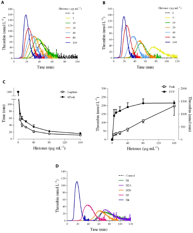
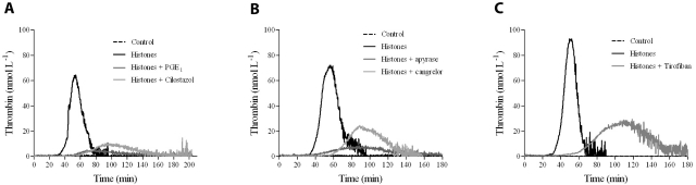
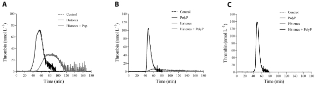
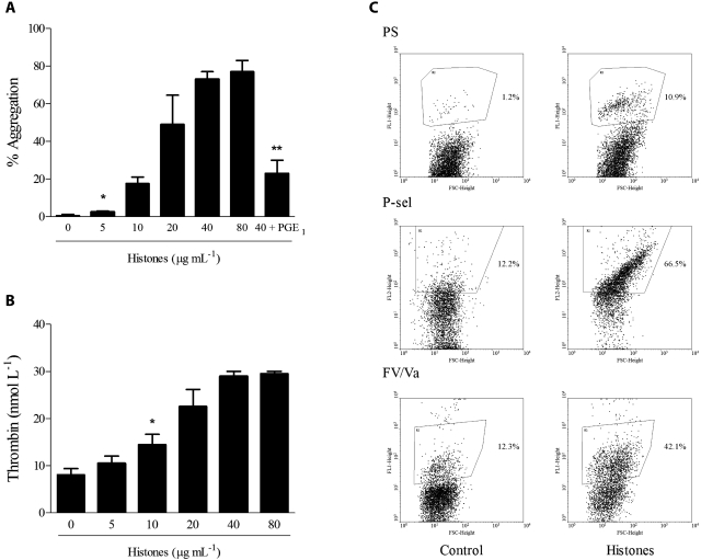
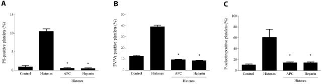
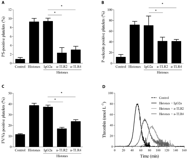
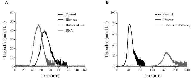
The release of histones from dying cells is associated with microvascular thrombosis and, because histones activate platelets, this could represent a possible pathogenic mechanism. In the present study, we assessed the influence of histones on the procoagulant potential of human platelets in platelet-rich plasma (PRP) and in purified systems. Histones dose-dependently enhanced thrombin generation in PRP in the absence of any trigger, as evaluated by calibrated automated thrombinography regardless of whether the contact phase was inhibited. Activation of coagulation required the presence of fully activatable platelets and was not ascribable to platelet tissue factor, whereas targeting polyphosphate with phosphatase reduced thrombin generation even when factor XII (FXII) was blocked or absent. In the presence of histones, purified polyphosphate was able to induce thrombin generation in plasma independently of FXII. In purified systems, histones induced platelet aggregation; P-selectin, phosphatidylserine, and FV/Va expression; and prothrombinase activity. Blocking platelet TLR2 and TLR4 with mAbs reduced the percentage of activated platelets and lowered the amount of thrombin generated in PRP. These data show that histone-activated platelets possess a procoagulant phenotype that drives plasma thrombin generation and suggest that TLR2 and TLR4 mediate the activation process.


Figures

Figure 1
Figure 1
Histones increase thrombin generation in human PRP. Thrombin generation was monitored in real time by CAT in the absence of any trigger. (A) Typical thrombograms in the presence of increasing concentrations of calf thymus histones (0-160 μg/mL) in recalcified citrated PRP. (B) Thrombograms in the presence of histones in recalcified CTI-inhibited PRP. Note that the control curve is flat. In both cases, a representative experiment of 4 performed with PRP from different donors is shown. The tails of the thrombograms have been cut for clarity in these and next graphs. (C) Changes in CAT parameters as a function of histone concentration in CTI-inhibited PRP. Lag time and time-to-peak (ttPeak) are shown on the left; thrombin peak and ETP on the right. Data are the means ± SD of 4 independent experiments. (D) Recalcified CTI-inhibited PRP was treated with single recombinant histones (20 μg/mL) and then thrombin generation was evaluated. A representative experiment of 3 performed with PRP from different donors is shown.
Figure 2
Figure 2
Platelet inhibitors reduce thrombin generation in histone-treated PRP. Recalcified CTI-inhibited PRP was treated with 10 μmol/L of PGE1 or 50 μmol/L of cilostazol (A), 10 U/mL of apyrase or 1 μmol/L of cangrelor (B), or 1.5 μmol/L of tirofiban (C) for 10 minutes at 37°C before stimulation with histones (40 μg/mL). Thrombin generation was then evaluated by CAT. A representative experiment of 4 performed with PRP from different donors is shown.
Figure 3
Figure 3
PolyP drives thrombin generation in histone-treated PRP. (A) Recalcified CTI-inhibited PRP was incubated with histones (40 μg/mL) in the presence of Psp (10 U/mL), and then thrombin generation was evaluated. Representative tracings of 3 experiments with PRP from different donors are shown. Recalcified CTI-inhibited PPP (B) and PPP treated with an anti-FXII antibody (150 μg/mL; C) were incubated with histones (40 μg/mL) and/or polyP (20 μg/mL), as described in “Methods,” and then thrombin generation was evaluated in the presence of liposomes. One representative experiment of 3 is shown.
Figure 4
Figure 4
Histones induce platelet aggregation, prothrombinase activity, PS exposure, and secretion. (A) Histones (0-80 μg/mL) were added to stirred gel-filtered human platelets, and aggregation was monitored for 10 minutes. Where indicated, gel-filtered platelets were pretreated with PGE1 (4 μmol/L) before the addition of histones (40 μg/mL). Data are reported as means of the maximum percentage of aggregation induced by histones ± SD of 4 independent experiments with gel-filtered platelets from different donors. *Lowest concentration producing significant aggregation (P < .05); **P < .05 compared with histones (40 μg/mL). (B) Prothrombinase activity of gel-filtered platelets stimulated with histones (0-80 μg/mL) for 15 minutes was determined after the addition of human FXa and prothrombin; the amount of thrombin generated was evaluated after 90 seconds by cleavage of the chromogenic substrate Spectrozyme TH. Data are means ± SD, n = 3. *Lowest concentration producing a significant increase (P < .05). (C) Typical flow cytometric analysis of PS exposure (top panel), P-selectin expression (middle panel), and FV/Va expression (bottom panel). Gel-filtered platelets stimulated with histones (40 μg/mL) for 15 minutes were analyzed by flow cytometry as detailed in “Methods.” Region 2 indicating a positive population for the antigen of interest was arbitrarily chosen.
Figure 5
Figure 5
APC and heparin abolish the platelet-activating potential of histones. Histones (40 μg/mL) pretreated with APC (6 μg/mL) or heparin (1 U/mL) were added to gel-filtered platelets as described in “Methods,” and then their platelet-activating potential was evaluated by measuring PS exposure (A), FV/Va (B), and P-selectin expression (C) by flow cytometry. Data are means ± SD, n = 3. *P < .05 compared with histones.
Figure 6
Figure 6
Histone-induced platelet activation is partially dependent on TLR2 and TLR4. Flow cytometric analysis of PS exposure (A), P-selectin expression (B), and FV/Va expression (C) in gel-filtered platelets stimulated with histones (40 μg/mL) with or without preincubation with mouse monoclonal anti–human TLR2 or TLR4 blocking Abs (50 μg/mL) or control mouse IgG2a for 20 minutes at RT. Data are means ± SD, n = 3. *P < .05. (D) Thrombin generation induced by histones (40 μg/mL) in CTI-inhibited PRP pretreated with neutralizing mAbs against TLR2, TLR4, or control Ab. Representative thrombograms of 3 experiments are shown.
Figure 7
Figure 7
Histone-DNA complexes increase thrombin generation in PRP. Thrombin generation tracings in recalcified CTI-inhibited PRP stimulated with DNA-histone complexes (1:1 weight ratio; A) or de-N-heparin histone complexes (0.5:1 weight ratio; B), as described in “Methods.” One representative tracing of 3 experiments is shown.

Comment in

References

    1. Jahr S, Hentze H, Englisch S, et al. DNA fragments in the blood plasma of cancer patients: quantitations and evidence for their origin from apoptotic and necrotic cells. Cancer Res. 2001;61(4):1659–1665. - PubMed
    1. Brinkmann V, Laube B, Abu Abed U, et al. Neutrophil extracellular traps kill bacteria. Science. 2004;303(5663):1532–1535. - PubMed
    1. von Köckritz-Blickwede M, Goldmann O, Thulin P, et al. Phagocytosis-independent antimicrobial activity of mast cells by means of extracellular trap formation. Blood. 2008;111(6):3070–3080. - PubMed
    1. Holdenrieder S, Stieber P. Clinical use of circulating nucleosomes. Crit Rev Clin Lab Sci. 2009;46(1):1–24. - PubMed
    1. Zeerleder S, Zwart B, Wuillemin WA, et al. Elevated nucleosome levels in systemic inflammation and sepsis. Crit Care Med. 2003;31(7):1947–1951. - PubMed

Publication types