. 2011 Jul 12;105(2):231-8.
doi: 10.1038/bjc.2011.226. Epub 2011 Jun 14.

CTGF is overexpressed in malignant melanoma and promotes cell invasion and migration

Affiliations

CTGF is overexpressed in malignant melanoma and promotes cell invasion and migration

S Braig et al. Br J Cancer. .

Abstract

Background: Malignant melanoma cells are known to have altered expression of growth factors compared with normal human melanocytes. These changes most likely favour tumour growth and progression, and influence tumour environment. The induction of transforming growth factor beta1, 2 and 3 as well as BMP4 and BMP7 expression in malignant melanoma has been reported before, whereas the expression of an important modulator of these molecules, connective tissue growth factor (CTGF), has not been investigated in melanomas until now.

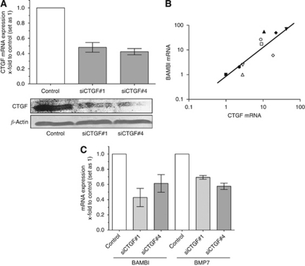
Methods: Expression of CTGF was analysed in melanoma cell lines and tissue samples by qRT-PCR and immunohistochemistry. To determine the regulation of CTGF expression in malignant melanoma, specific siRNA was used. Additionally, migration, invasion and attachment assays were carried out.

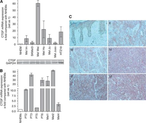
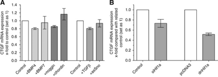
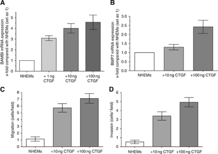
Results: We were able to demonstrate that CTGF expression is upregulated in nine melanoma cell lines and in primary and metastatic melanoma in situ. The transcription factor HIF-1α was revealed as a positive regulator for CTGF expression. Melanoma cells, in which CTGF expression is diminished, show a strong reduction of migratory and invasive properties when compared with controls. Further, treatment of normal human epidermal melanocytes with recombinant CTGF leads to an increase of migratory and invasive behaviour of these cells.

Conclusion: These results suggest that CTGF promotes melanoma cell invasion and migration and, therefore, has an important role in the progression of malignant melanoma.


Figures

Figure 1
Figure 1
Expression of CTGF in malignant melanoma. The expression of CTGF mRNA was analysed by quantitative RT–PCR (A) and western blot in NHEMs and in the melanoma cell lines Mel Im, Mel Ju, Mel Juso, Mel Ho, Mel Wei, SK Mel 3 and HTZ19d. (B) Further, CTGF mRNA expression was determined in tissue samples of primary melanoma (PT2–PT6) and melanoma metastasis (Met1–Met4) by quantitative RT–PCR. (C) Connective tissue growth factor protein expression was analysed by immunohistochemistry in skin (I), primary melanoma (II and III) and metastasis of malignant melanoma (IV, V and VI). Strong expression of CTGF protein was detected in primary melanoma and metastases as shown by red AEC staining (200-fold magnification). Bars show the mean±s.d. of three independent experiments, measurements were performed in triplicates. The colour reproduction of this figure is available at the British Journal of Cancer online.
Figure 2
Figure 2
Analysis of regulation of CTGF by HIF-1α. (A) Mel Im cells were treated with recombinant BMP4 or BMP7, respectively. Quantitative RT–PCR analysis revealed no difference in CTGF mRNA expression levels compared with untreated cells. Inhibition of BMP signalling by treatment of the cells with noggin and chordin had no effect on CTGF mRNA expression. Neither treatment of melanoma cells with TGFβ1 nor transfection of Mel Im cells with an antisense Sno construct influenced mRNA expression of CTGF. (B) Transient transfection of Mel Im cells with siRNA against HIF-1α resulted in diminished CTGF mRNA expression. Moreover, transfection of Mel Im cells with a dominant-negative HIF-1α construct strongly reduced CTGF gene expression compared with cells treated with pcDNA3 control vector. Bars show the mean±s.d. of three independent experiments, measurements were performed in triplicates.
Figure 3
Figure 3
Connective tissue growth factor regulates expression of BAMBI and BMP7. (A) Reduction of CTGF expression was achieved by transient transfection of Mel Im cells with siRNAs against CTGF (siCTGF#1 and siCTGF#4). Transfection with scrambled siRNA served as control. Strong reduction of CTGF expression was observed after 48 h in the melanoma cell line Mel Im on mRNA and protein level. (B) Correlation of CTGF and BAMBI mRNA expression profiles in melanoma cell lines. Each symbol represents one melanoma cell line. (C) Transient transfection of melanoma Mel Im cells with both siRNAs against CTGF resulted in reduced expression of BAMBI and BMP7 mRNA levels compared with control cells, as determined by qRTPCR. Bars show the mean±s.d. of three independent experiments, measurements were performed in triplicates.
Figure 4
Figure 4
Effect of reduction of CTGF expression. (A) Migration and invasion assays using the Boyden Chamber model revealed a significant reduction of the migratory and invasive potential after transient downregulation of CTGF expression, whereas attachment of the cells (B) was not significantly changed. Bars show the mean±s.d. of three independent experiments, measurements were performed in triplicates. **P<0.01; ***P<0.001.
Figure 5
Figure 5
Treatment of NHEMs with CTGF results in increased expression of BAMBI and BMP7 and enhanced migratory and invasive potential. (A and B) Normal human epidermal melanocytes were treated with increasing concentrations of recombinant CTGF for 24 h. Quantitative RT–PCR revealed enhanced expression of BAMBI (A) and BMP7 mRNA (B) levels in NHEMs incubated with CTGF compared with control cells. (C) Connective tissue growth factor-treated cells showed strongly increased migratory potential of up to 700% when compared with untreated melanocytes. (D) In addition, invasive potential of NHEMs was dose dependently enhanced up to 500% after treatment with recombinant CTGF compared with control cells. Bars show the mean±s.d. of three independent experiments, measurements were performed in triplicates.

References

    1. Babic AM, Chen CC, Lau LF (1999) Fisp12/mouse connective tissue growth factor mediates endothelial cell adhesion and migration through integrin alphavbeta3, promotes endothelial cell survival, and induces angiogenesis in vivo. Mol Cell Biol 19: 2958–2966 - PMC - PubMed
    1. Barcellos-Hoff MH, Akhurst RJ (2009) Transforming growth factor-beta in breast cancer: too much, too late. Breast Cancer Res 11: 202. - PMC - PubMed
    1. Bellam N, Pasche B (2010) Tgf-beta signaling alterations and colon cancer. Cancer Treat Res 155: 85–103 - PubMed
    1. Bradham DM, Igarashi A, Potter RL, Grotendorst GR (1991) Connective tissue growth factor: a cysteine-rich mitogen secreted by human vascular endothelial cells is related to the SRC-induced immediate early gene product CEF-10. J Cell Biol 114: 1285–1294 - PMC - PubMed
    1. Chen XM, Qi W, Pollock CA (2009) CTGF and chronic kidney fibrosis. Front Biosci (Schol Ed) 1: 132–141 - PubMed

MeSH terms