

Imaging of melanoma: usefulness of ultrasonography before and after contrast injection for diagnosis and early evaluation of treatment
- PMID: 21673868
- PMCID: PMC3108283
- DOI: 10.2147/CCID.S13499
Imaging of melanoma: usefulness of ultrasonography before and after contrast injection for diagnosis and early evaluation of treatment
Abstract
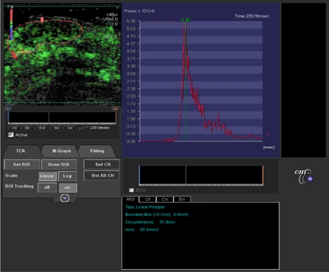
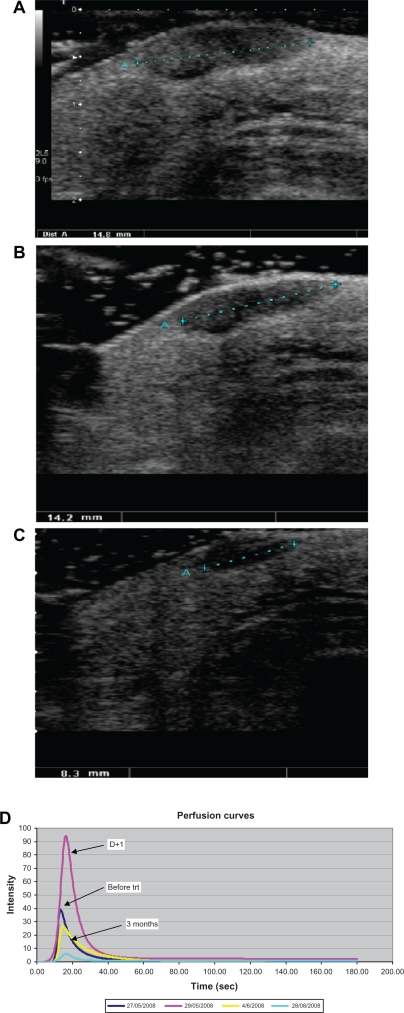
High-frequency ultrasound (8-14 MHz) is routinely used to display cutaneous melanomas. Maximum thickness measurement (Breslow index) has been shown to be well correlated to histologic findings for lesions of more than 0.75 mm. Some morphological criteria (strong delineation, hypoechoic texture, homogeneity) have been reported to help differentiate between malignant and benign pigmented blue lesions, but remain insufficient. Vascular ultrasound analysis using Doppler mode provides additional information and showed good specificity for malignancy (90%-100%), but variable sensitivity (34%-100%). Recent advances in ultrasound imaging allow functional evaluation. Likewise, dynamic contrast-enhanced ultrasound using contrast medium injection and specific perfusion and quantification software showed promising results in clinical and preclinical trials for early prediction of tumor response to target treatments.
Keywords: dynamic contrast-enhanced ultrasound; melanoma; ultrasound.
Figures
References
-
- Kraus W, Schramm P, Hoede N. First experiences with a high resolution ultrasonic scanner in the diagnosis of malignant melanomas. Arch Dermato Res. 1983;275:235–238. - PubMed
-
- Dines K, Sheets P, Bink J, et al. High-frequency ultrasonic imaging of skin: Experimental results. Ultrason Imaging. 1984;6:408–434. - PubMed
-
- Hoffman K, Jung J, El Gammal S, Altmeyer P. Malignant melanoma in 20-MHz B scan US. Dermatology. 1992;185:49–55. - PubMed
-
- Fornage B, McGavran M, Duvic M, Waldron C. Imaging of the skin with 20 MHz sonography. Radiology. 1993;189:69–79. - PubMed
LinkOut - more resources
Full Text Sources
