. 2011;12(3):1595-604.
doi: 10.3390/ijms12031595. Epub 2011 Mar 1.

Cancer stem-like cells enriched in Panc-1 spheres possess increased migration ability and resistance to gemcitabine

Affiliations

Cancer stem-like cells enriched in Panc-1 spheres possess increased migration ability and resistance to gemcitabine

Tao Yin et al. Int J Mol Sci. 2011.

Abstract

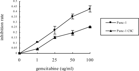
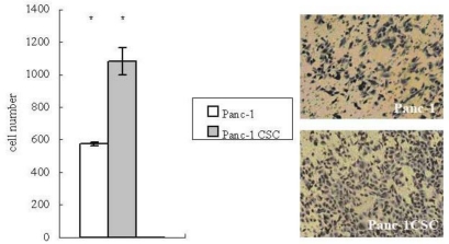
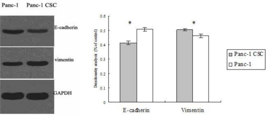
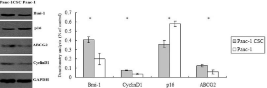
Pancreatic cancer is one of the most lethal malignancies with poor prognosis. Previously, we found that a subpopulation of cancer stem cells (CSCs) in the Panc-1 pancreatic cancer cell line could propagate to form spheres. Here we characterized the malignant phenotypes of the pancreatic cancer stem CD44+/CD24+ cells, which were enriched under sphere forming conditions as analyzed by flow cytometry. These cells demonstrated increased resistance to gemcitabine and increased migration ability. Moreover, these cells exhibited epithelial to mesenchymal transition characterized by a decreased level of the epithelial marker E-cadherin and an increased level of the mesenchymal marker vimentin. Notably, abnormal expression of Bmi-1, ABCG2, Cyclin D1 and p16 were found in Panc-1 CSCs. Our results suggest that targeted inhibition of CSCs represents a novel therapeutic approach to overcome chemoresistance and metastasis of pancreatic cancer.

Keywords: Bmi-1; chemoresistance; invasion; pancreatic cancer; tumorigenesis.


Figures

Figure 1.
Figure 1.
The Panc-1 cell cancer stem cell (CSC) subpopulation (CD44+/CD24+) was enriched under sphere-forming conditions. The right upper quadrant (Q2-2) indicates the distribution of the CD44+/CD24+ subgroup of cells as analyzed by flow cytometry. The experiment was repeated 5 times and the data are expressed as mean ± standard deviation, statistically significant differences were determined by Student’s t test, the difference between the Panc-1 group and the Panc-1 CSC group was significant, p < 0.05.
Figure 2.
Figure 2.
Panc-1 CSCs have increased chemoresistance to gemcitabine. Panc-1 CSCs or Panc-1 cells were treated with various dosages of gemcitabine for 48 h. Cell viability was determined by MTT assay. Panc-1 CSCs showed increased resistance to gemcitabine compared to Panc-1 cells cultured in monolayer.
Figure 3.
Figure 3.
Panc-1 CSCs have increased migration ability. The migration of Panc-1 CSCs or Panc-1 cells was determined by transwell migration assay. After the filter was fixed and stained, the number of cells migrated to the undersurface of the filter was compared. Left side: Panc-1 CSCs had increased migration ability compared to Panc-1 cells, (40× fold). * p < 0.05. Right side: images of Panc-1 cells (top) and Panc-1 CSCs (bottom).
Figure 4.
Figure 4.
Panc-1 CSCs demonstrate a phenotype suggestive of epithelial to mesenchymal transition (EMT). The expression levels of E-cadherin and vimentin in Panc-1 CSCs and Panc-1 cells were determined by Western blot analysis (left). Densitometry analysis revealed the differences between Panc-1 CSCs and Panc-1 cells (right). * p < 0.05.
Figure 5.
Figure 5.
Abnormal expression of Bmi-1, ABCG2, Cyclin D1 and p16 in Panc-1 CSCs. The levels of Bmi-1, ABCG2 and p16 in Panc-1 CSCs and Panc-1 cells were determined by Western blot analysis and normalized to GAPDH (left). Densitometry analysis revealed the differences between Panc-1 CSCs and Panc-1 cells. * p < 0.05.

References

    1. Jemal A, Tiwari RC, Murray T. Cancer statistics, 2004. CA Cancer J. Clin. 2004;54:8–29. - PubMed
    1. Polyak K, Hahn WC. Roots and stems, stem cells in cancer. Nat. Med. 2006;12:296–300. - PubMed
    1. Li F, Tiede B, Massagué J, Kang Y. Beyond tumorigenesis, cancer stem cells in metastasis. Cell Res. 2007;17:3–14. - PubMed
    1. Kakarala M, Wicha MS. Implications of the cancer stem-cell hypothesis for breast cancer prevention and therapy. J. Clin. Oncol. 2008;26:2813–2820. - PMC - PubMed
    1. Singh SK, Clarke ID, Terasaki M, Bonn VE, Hawkins C, Squire J, Dirks PB. Identification of a cancer stem cell in human brain tumors. Cancer Res. 2003;63:5821–5828. - PubMed

Publication types

MeSH terms