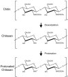
Chitin-based materials in tissue engineering: applications in soft tissue and epithelial organ
- PMID: 21673932
- PMCID: PMC3111643
- DOI: 10.3390/ijms12031936
Chitin-based materials in tissue engineering: applications in soft tissue and epithelial organ
Abstract
Chitin-based materials and their derivatives are receiving increased attention in tissue engineering because of their unique and appealing biological properties. In this review, we summarize the biomedical potential of chitin-based materials, specifically focusing on chitosan, in tissue engineering approaches for epithelial and soft tissues. Both types of tissues play an important role in supporting anatomical structures and physiological functions. Because of the attractive features of chitin-based materials, many characteristics beneficial to tissue regeneration including the preservation of cellular phenotype, binding and enhancement of bioactive factors, control of gene expression, and synthesis and deposition of tissue-specific extracellular matrix are well-regulated by chitin-based scaffolds. These scaffolds can be used in repairing body surface linings, reconstructing tissue structures, regenerating connective tissue, and supporting nerve and vascular growth and connection. The novel use of these scaffolds in promoting the regeneration of various tissues originating from the epithelium and soft tissue demonstrates that these chitin-based materials have versatile properties and functionality and serve as promising substrates for a great number of future applications.
Keywords: chitin; chitosan; epithelium; regeneration; soft tissue; tissue engineering.
Figures


References
-
- Kumar MN, Muzzarelli RA, Muzzarelli C, Sashiwa H, Domb AJ. Chitosan chemistry and pharmaceutical perspectives. Chem. Rev. 2004;104:6017–6084. - PubMed
-
- Felt O, Furrer P, Mayer JM, Plazonnet B, Buri P, Gurny R. Topical use of chitosan in ophtalmology: Tolerance assessment and evaluation of precorneal retention. Int. J. Pharm. 1999;180:185–193. - PubMed
-
- Kim IY, Seo SJ, Moon HS, Yoo MK, Park IY, Kim BC, Cho CS. Chitosan and its derivatives for tissue engineering applications. Biotechnol. Adv. 2008;26:1–21. - PubMed
-
- Chung YC, Chen CY. Antibacterial characteristics and activity of acid-soluble chitosan. Bioresour. Technol. 2008;99:2806–2814. - PubMed
Publication types
MeSH terms
Substances
LinkOut - more resources
Full Text Sources
Other Literature Sources

