

Chaperoning roles of macromolecules interacting with proteins in vivo
- PMID: 21673934
- PMCID: PMC3111645
- DOI: 10.3390/ijms12031979
Chaperoning roles of macromolecules interacting with proteins in vivo
Abstract
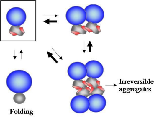
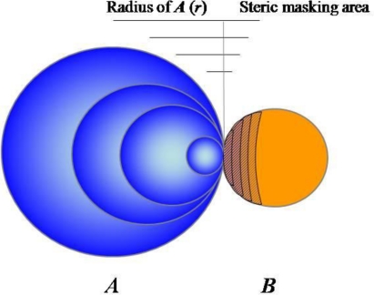
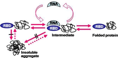
The principles obtained from studies on molecular chaperones have provided explanations for the assisted protein folding in vivo. However, the majority of proteins can fold without the assistance of the known molecular chaperones, and little attention has been paid to the potential chaperoning roles of other macromolecules. During protein biogenesis and folding, newly synthesized polypeptide chains interact with a variety of macromolecules, including ribosomes, RNAs, cytoskeleton, lipid bilayer, proteolytic system, etc. In general, the hydrophobic interactions between molecular chaperones and their substrates have been widely believed to be mainly responsible for the substrate stabilization against aggregation. Emerging evidence now indicates that other features of macromolecules such as their surface charges, probably resulting in electrostatic repulsions, and steric hindrance, could play a key role in the stabilization of their linked proteins against aggregation. Such stabilizing mechanisms are expected to give new insights into our understanding of the chaperoning functions for de novo protein folding. In this review, we will discuss the possible chaperoning roles of these macromolecules in de novo folding, based on their charge and steric features.
Keywords: aggregation; de novo folding; hydrophobic interactions; macromolecules; molecular chaperones; stabilization; steric hindrance; surface charges.
Figures
References
-
- Bukau B, Horwich AL. The Hsp70 and Hsp60 chaperone machines. Cell. 1998;92:351–366. - PubMed
-
- Hartl FU, Hayer-Hartl M. Molecular chaperones in the cytosol: from nascent chain to folded protein. Science. 2002;295:1852–1858. - PubMed
-
- Anfinsen CB. Principles that govern the folding of protein chains. Science. 1973;181:223–230. - PubMed
Publication types
MeSH terms
Substances
LinkOut - more resources
Full Text Sources
