. 2011;6(6):e20758.
doi: 10.1371/journal.pone.0020758. Epub 2011 Jun 2.

Transmigration of melanoma cells through the blood-brain barrier: role of endothelial tight junctions and melanoma-released serine proteases

Affiliations

Transmigration of melanoma cells through the blood-brain barrier: role of endothelial tight junctions and melanoma-released serine proteases

Csilla Fazakas et al. PLoS One. 2011.

Abstract

Malignant melanoma represents the third common cause of brain metastasis, having the highest propensity to metastasize to the brain of all primary neoplasms in adults. Since the central nervous system lacks a lymphatic system, the only possibility for melanoma cells to reach the brain is via the blood stream and the blood-brain barrier. Despite the great clinical importance, mechanisms of transmigration of melanoma cells through the blood-brain barrier are incompletely understood. In order to investigate this question we have used an in vitro experimental setup based on the culture of cerebral endothelial cells (CECs) and the A2058 and B16/F10 melanoma cell lines, respectively. Melanoma cells were able to adhere to confluent brain endothelial cells, a process followed by elimination of protrusions and transmigration from the luminal to the basolateral side of the endothelial monolayers. The transmigration process of certain cells was accelerated when they were able to use the routes preformed by previously transmigrated melanoma cells. After migrating through the endothelial monolayer several melanoma cells continued their movement beneath the endothelial cell layer. Melanoma cells coming in contact with brain endothelial cells disrupted the tight and adherens junctions of CECs and used (at least partially) the paracellular transmigration pathway. During this process melanoma cells produced and released large amounts of proteolytic enzymes, mainly gelatinolytic serine proteases, including seprase. The serine protease inhibitor Pefabloc® was able to decrease to 44-55% the number of melanoma cells migrating through CECs. Our results suggest that release of serine proteases by melanoma cells and disintegration of the interendothelial junctional complex are main steps in the formation of brain metastases in malignant melanoma.


Conflict of interest statement

Competing Interests: The authors have declared that no competing interests exist.

Figures

Figure 1
Figure 1. Adhesion of melanoma cells to brain endothelial cells.
Fluorescently labeled melanoma cells (A2058 or B16/F10) (2.5·104/cm2) were plated onto confluent CECs (RBEC or D3) and left for different time intervals. After washing of non-adherent cells, attached melanoma cells were counted.
Figure 2
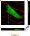
Figure 2. Migration of melanoma cells from the apical to the basolateral side of CECs.
RBECs were grown on glass coverslips. A2058 melanoma cells labeled with OG were plated onto confluent endothelial monolayers. After 5 h cells were washed, fixed and tight junctions were stained with anti-ZO-1 antibody (red). Samples were analysed by confocal laser scanning microscopy. A: xy-stack at the level of the green lines on B and C. B, C: z-stacks along the vertical and horizontal line, respectively on A.
Figure 3
Figure 3. Changes in the transendothelial electrical resistance in the presence of melanoma cells.
RBECs were grown on semipermeable filters with 0.4 µm pore size. TEER was followed using the CellZscope system. N = 2, * = P<0.05 as assessed by ANOVA and Bonferroni's post hoc test.
Figure 4
Figure 4. Induction of endothelial apoptosis by melanoma cells.
RBECs were cultured on glass coverslips. A2058 melanoma cells labeled with CellTracker™ Blue were plated onto confluent endothelial monolayers. After 24 h cells were washed and fixed. Tight junctions were stained with anti-ZO-1 antibody (marker of endothelial cells), while apoptotic cells were visualized using anti-cleaved caspase-3 antibody. Apoptotic endothelial cells were counted. N = 2, * = P<0.05 as assessed by Student's t-test.
Figure 5
Figure 5. Adhesion of melanoma cells to interendothelial junctions.
RBECs were grown on 8 µm pore size filter inserts. After reaching confluency A2058 cells were plated into the upper chamber and left for 5 h. Samples were fixed and 60–70 nm sections were prepared for electron microscopy. Arrow indicates interendothelial cell contact site. M = melanoma cell, E = endothelial cell. Scale bar = 10 µm.
Figure 6
Figure 6. Disruption of interendothelial tight junctions induced by melanoma cells.
Fluorescently labeled (A, B: CellTracker™ Blue, C, D: OG) melanoma cells (A, B, C: A2058 or D: B16/F10) were plated onto confluent brain endothelial cells (RBEC) and left for 2 (A) or 5 h (B–D). After washing of non-attached melanoma cells samples were fixed and stained for claudin-5, ZO-1 or occludin. Arrows indicate sites of disrupted junctions. Scale bar = 50 µm.
Figure 7
Figure 7. Changes in the total amount of endothelial junctional proteins in the presence of melanoma cells or melanoma-released factors.
Melanoma cells or melanoma conditioned media were placed onto confluent brain endothelial cells (A, C: RBEC; B, D: D3) and the amount of claudin-5 (A, B) or occludin (C, D) was assessed by Western-blot analysis.
Figure 8
Figure 8. Role of gelatinolytic serine proteases produced by melanoma cells.
A, B: Melanoma cells were plated onto confluent monolayers of cerebral endothelial cells or into empty culture dishes in serum-free medium in the presence or absence of E64 or Pefabloc® and left for 5 h. Culture media were collected and cells were lysed in Triton X-114 containing buffer. Samples were electrophoresed in non-denaturing conditions and the gels were incubated in EDTA-containing buffer for 2 days. Proteolytic bands of culture media (A) or cell lysates (B) were visualized by Coomassie blue staining. C, D: RBEC were grown until confluency on 8 µm pore size filter inserts. Fluorescently labeled melanoma cells (C: A2058, D: B16/F10) were plated into the upper chamber in the presence or absence of Pefabloc® and left for 5 h. Cells from the upper chamber were removed using a cotton swab, and melanoma cells migrated through the endothelial cell layer and the pores of the filter were counted. N = 3, * = P<0.05 as assessed by Student's t-test.
Figure 9
Figure 9. Role of seprase in the transmigration of A2058 cells through brain endothelial cells.
A: Seprase was silenced in A2058 cells (fold change = 0.09 compared to scrambled RNA-transfected cells, determined by real-time PCR). Melanoma cells were plated onto confluent monolayers of cerebral endothelial cells or into empty culture dishes in serum-free medium and left for 5 h. Cells were lysed in Triton X-114 containing buffer. Zymography was performed in the presence of EDTA. Arrow indicates seprase. B: Seprase-silenced or scrambled RNA-transfected cells were plated onto confluent RBECs grown on 8 µm pore size filter inserts. Transmigration assay was performed and melanoma cells reaching the bottom of the filter inserts were counted. N = 3, * = P<0.05 as assessed by Student's t-test.

References

    1. Denkins Y, Reiland J, Roy M, Sinnappah-Kang ND, Galjour J, et al. Brain metastases in melanoma: roles of neurotrophins. Neuro Oncol. 2004;6:154–165. - PMC - PubMed
    1. Sloan AE, Nock CJ, Einstein DB. Diagnosis and treatment of melanoma brain metastasis: a literature review. Cancer Control. 2009;16:248–255. - PubMed
    1. Abbott NJ, Patabendige AA, Dolman DE, Yusof SR, Begley DJ. Structure and function of the blood-brain barrier. Neurobiol Dis. 2010;37:13–25. - PubMed
    1. Bauer HC, Traweger A, Zweimueller-Mayer J, Lehner C, Tempfer H, et al. New aspects of the molecular constituents of tissue barriers. J Neural Transm. 2011;118:7–21. - PubMed
    1. Qi J, Chen N, Wang J, Siu, CH Transendothelial migration of melanoma cells involves N-cadherin-mediated adhesion and activation of the beta-catenin signaling pathway. Mol Biol Cell. 2005;16:4386–4397. - PMC - PubMed

Publication types

Substances