

Tract-based magnetic resonance spectroscopy of the cingulum bundles at 7 T
- PMID: 21674690
- PMCID: PMC6870248
- DOI: 10.1002/hbm.21302
Tract-based magnetic resonance spectroscopy of the cingulum bundles at 7 T
Abstract
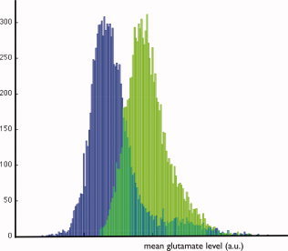
The cingulum bundle is a white matter fiber bundle in the human brain that is believed to be implicated in various neurological and psychiatric diseases. Subtle disease-related differences in metabolite concentrations in the cingulum tracts that may underlie these diseases may be detected using MR spectroscopic information. However, to date, limited signal to noise and lack of spatial resolution have prevented a reliable and reproducible measurement of metabolites in the cingulum bundle in vivo. Here we propose a new method that combines MR spectroscopic imaging at 7 T with fiber tracking to select only those MR spectroscopy voxels that are actually part of the cingulum bundles. The spectra of the selected spectroscopy voxels are processed per voxel and then combined yielding one spectrum at high spectral resolution for each cingulum bundle. In this way sensitivity is increased, as large parts of the cingulum are included while partial volume effects with both gray matter and white matter from other tracts is kept to a minimum. Three healthy volunteers were scanned to assess the feasibility of the method. For all three healthy volunteers spectra for the left and right cingulum tracts were computed, partial volume fractions calculated and metabolite fractions were quantified yielding similar results suggesting that tract-based MR spectroscopy allows us to study metabolic concentrations of individual white matter fiber bundles with high sensitivity and high specificity.
Copyright © 2011 Wiley-Liss, Inc.
Figures


References
-
- Andersson JL, Skare S ( 2002): A model‐based method for retrospective correction of geometric distortions in diffusion‐weighted EPI. Neuroimage 16: 177–199. - PubMed
-
- Andersson JL, Skare S, Ashburner J ( 2003): How to correct susceptibility distortions in spin‐echo echo‐planar images: Application to diffusion tensor imaging. Neuroimage 20: 870–888. - PubMed
-
- Basser PJ, Mattiello J, LeBihan D ( 1994): Estimation of the effective self‐diffusion tensor from the NMR spin echo. J Magn Reson B 103: 247–254. - PubMed
-
- Bloemen OJ, Deeley Q, Sundram F, Daly EM, Barker GJ, Jones DK, van Amelsvoort TA, Schmitz N, Robertson D, Murphy KC, Murphy DG ( 2010): White matter integrity in Asperger syndrome: A preliminary diffusion tensor magnetic resonance imaging study in adults. Autism Res 3: 203–213. - PubMed
-
- Boer VO, Siero JC, Hoogduin H, van Gorp JS, Luijten PR, Klomp DW ( 2011): High‐field MRS of the human brain at short TE and TR NMR Biomed. DOI:10.1002/nbm.1660. - PubMed
MeSH terms
LinkOut - more resources
Full Text Sources
