

Mechanized silica nanoparticles: a new frontier in theranostic nanomedicine
- PMID: 21675720
- PMCID: PMC3196789
- DOI: 10.1021/ar200018x
Mechanized silica nanoparticles: a new frontier in theranostic nanomedicine
Abstract
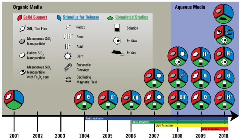
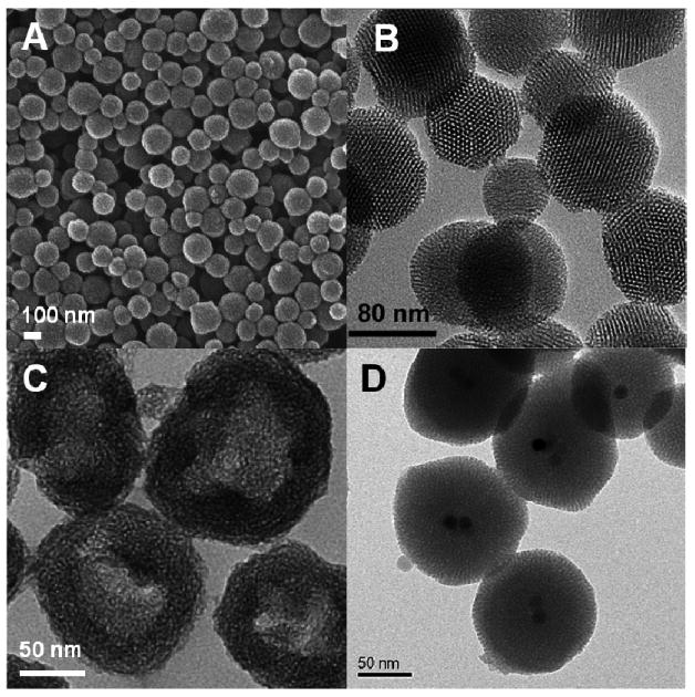
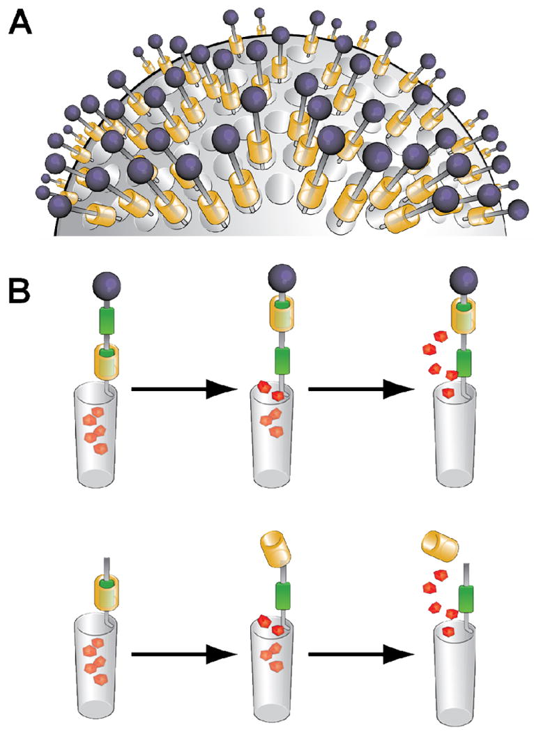
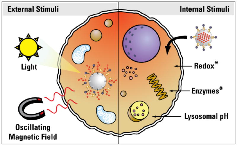
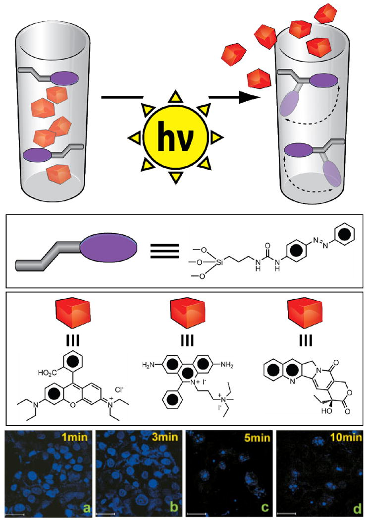
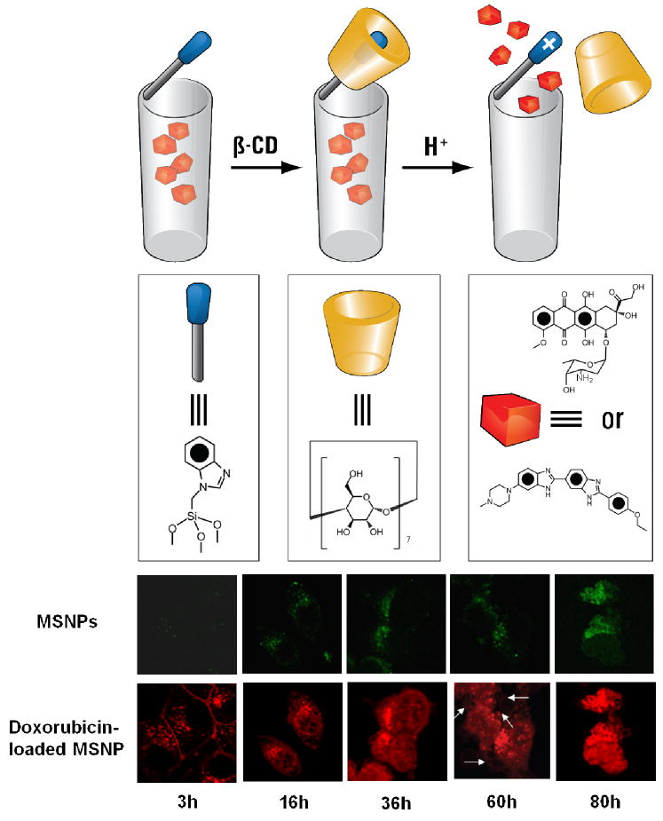
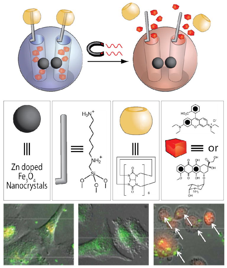
Medicine can benefit significantly from advances in nanotechnology because nanoscale assemblies promise to improve on previously established therapeutic and diagnostic regimes. Over the past decade, the use of delivery platforms has attracted attention as researchers shift their focus toward new ways to deliver therapeutic and/or diagnostic agents and away from the development of new drug candidates. Metaphorically, the use of delivery platforms in medicine can be viewed as the "bow-and-arrow" approach, where the drugs are the arrows and the delivery vehicles are the bows. Even if one possesses the best arrows that money can buy, they will not be useful if one does not have the appropriate bow to deliver the arrows to their intended location. Currently, many strategies exist for the delivery of bioactive agents within living tissue. Polymers, dendrimers, micelles, vesicles, and nanoparticles have all been investigated for their use as possible delivery vehicles. With the growth of nanomedicine, one can envisage the possibility of fabricating a theranostic vector that could release powerful therapeutics and diagnostic markers simultaneously and selectively to diseased tissue. In our design of more robust theranostic delivery systems, we have focused our attention on using mesoporous silica nanoparticles (SNPs). The payload "cargo" molecules can be stored within this robust domain, which is stable to a wide range of chemical conditions. This stability allows SNPs to be functionalized with stimulus-responsive mechanically interlocked molecules (MIMs) in the shape of bistable rotaxanes and psuedorotaxanes to yield mechanized silica nanoparticles (MSNPs). In this Account, we chronicle the evolution of various MSNPs, which came about as a result of our decade-long collaboration, and discuss advances in the synthesis of novel hybrid SNPs and the various MIMs which have been attached to their surfaces. These MIMs can be designed in such a way that they either change shape or shed off some of their parts in response to a specific stimulus, such as changes in redox potential, alterations in pH, irradiation with light, or the application of an oscillating magnetic field, allowing a theranostic payload to be released from the nanopores to a precise location at the appropiate time. We have also shown that these integrated systems can operate not only within cells, but also in live animals in response to pre-existing biological triggers. Recognizing that the theranostics of the future could offer a fresh approach to the treatment of degenerative diseases including cancer, we aim to start moving out of the chemical domain and into the biological one. Some MSNPs are already being tested in biological systems.
Figures

References
-
- Cotí KK, Belowich ME, Liong M, Ambrogio MW, Lau YA, Khatib HA, Zink JI, Khashab NM, Stoddart JF. Mechanised Nanoparticles for Drug Delivery. Nanoscale. 2009;1:16–39. - PubMed
-
- Klajn R, Stoddart JF, Grzybowski BA. Nanoparticles Functionalised with Reversible Molecular and Supramolecular Switches. Chem Soc Rev. 2010;39:2203–2237. - PubMed
-
- Mal NK, Fujiwara M, Tanaka Y. Photocontrolled Reversible Release of Guest Molecules from Coumarin-Modified Mesoporous Silica. Nature. 2003;421:350–353. - PubMed
-
- Liu R, Zhao X, Wu T, Feng P. Tunable Redox-Responsive Hybrid Nanogated Ensembles. J Am Chem Soc. 2008;130:14418–14419. - PubMed
-
- Park C, Kim H, Kim S, Kim C. Enzyme Responsive Nanocontainers with Cyclodextrin Gatekeepers and Synergistic Effects in Release of Guests. J Am Chem Soc. 2009;131:16614–16615. - PubMed
Publication types
MeSH terms
Substances
Grants and funding
LinkOut - more resources
Full Text Sources
Other Literature Sources
Miscellaneous
