. 2011 Aug;278(16):2913-26.
doi: 10.1111/j.1742-4658.2011.08216.x. Epub 2011 Jul 1.

An alternative isomerohydrolase in the retinal Müller cells of a cone-dominant species

Affiliations

An alternative isomerohydrolase in the retinal Müller cells of a cone-dominant species

Yusuke Takahashi et al. FEBS J. 2011 Aug.

Abstract

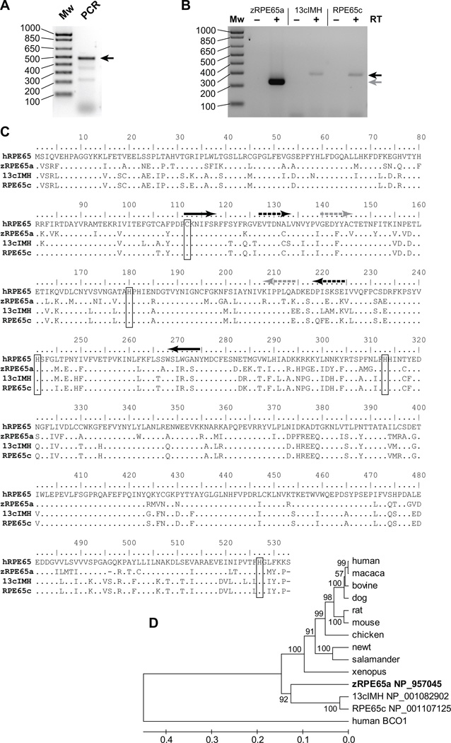
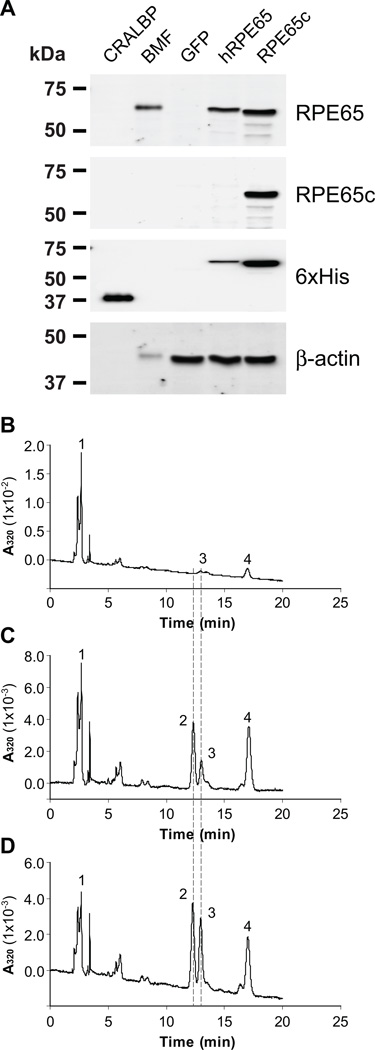
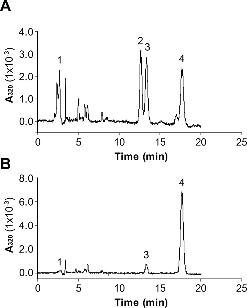
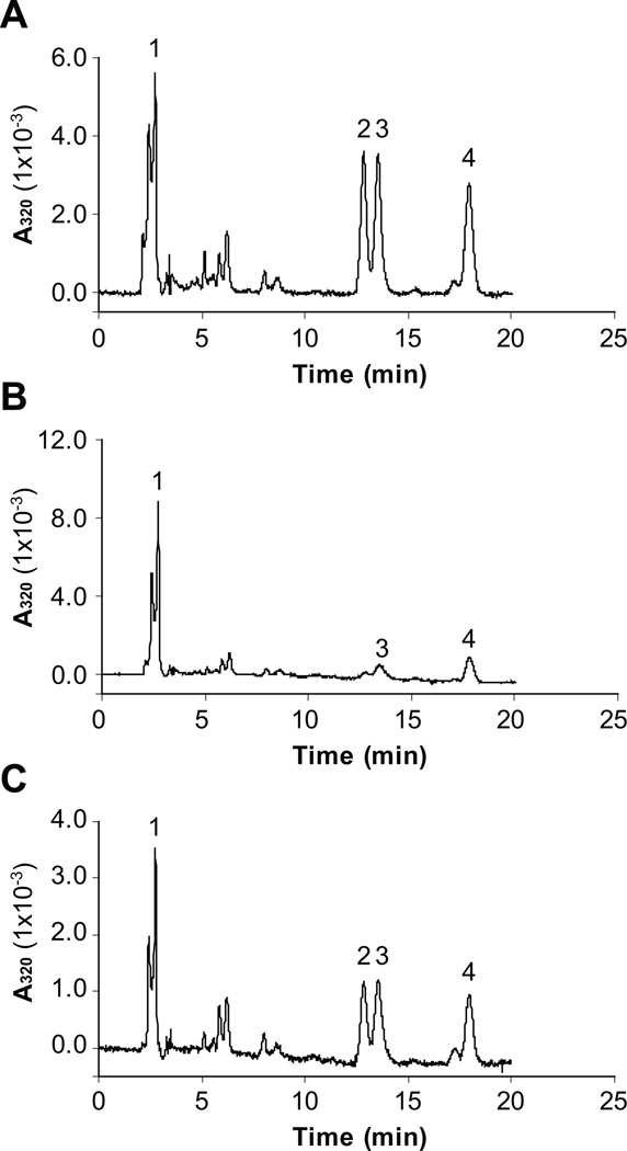
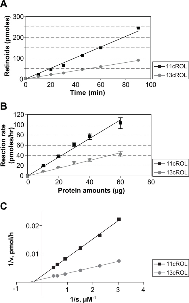
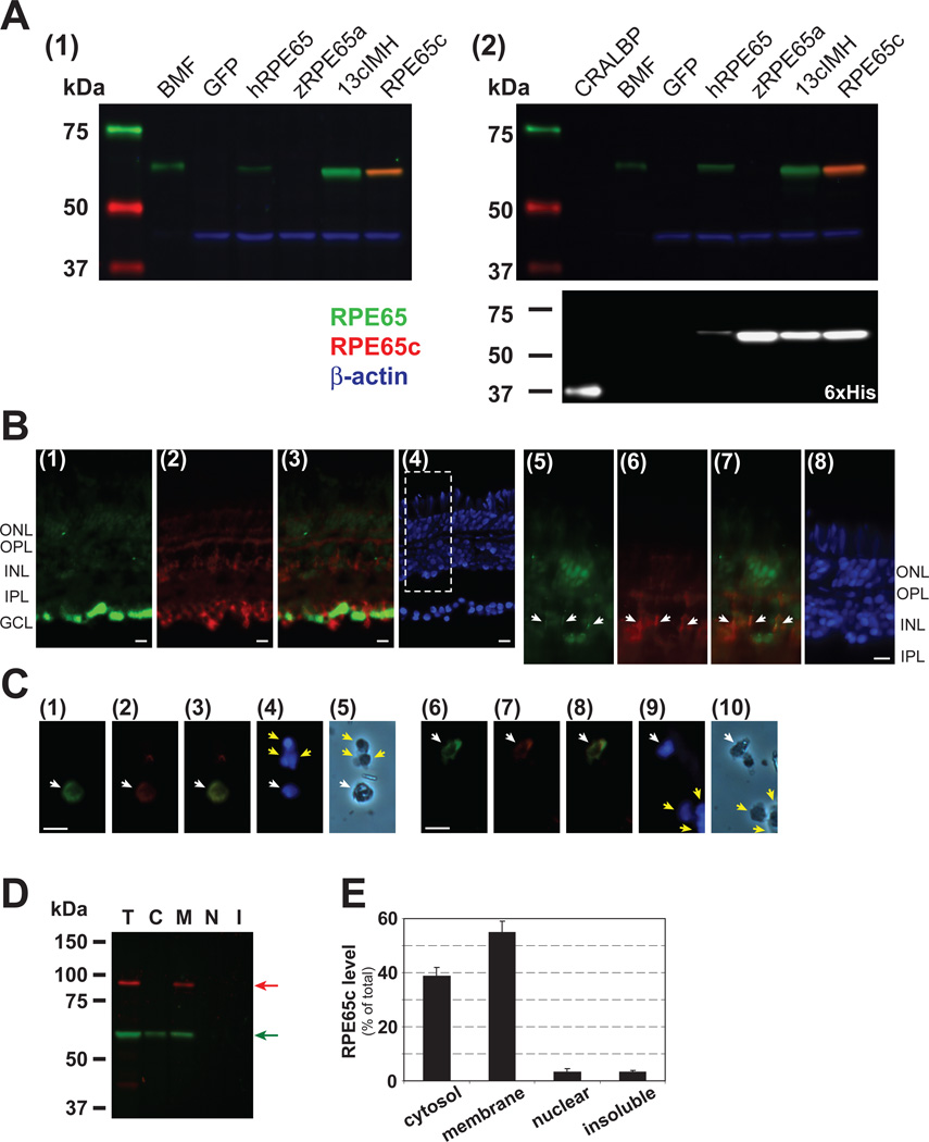
Cone photoreceptors have faster light responses than rods and a higher demand for 11-cis retinal (11cRAL), the chromophore of visual pigments. RPE65 is the isomerohydrolase in the retinal pigment epithelium (RPE) that converts all-trans retinyl ester to 11-cis retinol, a key step in the visual cycle for regenerating 11cRAL. Accumulating evidence suggests that cone-dominant species express an alternative isomerase, likely in retinal Müller cells, to meet the high demand for the chromophore by cones. In the present study, we describe the identification and characterization of a novel isomerohydrolase, RPE65c, from the cone-dominant zebrafish retina. RPE65c shares 78% amino acid sequence identity with RPE-specific zebrafish RPE65a (orthologue of human RPE65) and retains all of the known key residues for the enzymatic activity of RPE65. Similar to the other RPE-specific RPE65, RPE65c was present in both the membrane and cytosolic fractions, used all-trans retinyl ester as its substrate and required iron for its enzymatic activity. However, immunohistochemistry detected RPE65c in the inner retina, including Müller cells, but not in the RPE. Furthermore, double-immunostaining of dissociated retinal cells using antibodies for RPE65c and glutamine synthetase (a Müller cell marker), showed that RPE65c co-localized with the Müller cell marker. These results suggest that RPE65c is the alternative isomerohydrolase in the intra-retinal visual cycle, providing 11cRAL to cone photoreceptors in cone-dominant species. Identification of an alternative visual cycle will contribute to the understanding of the functional differences of rod and cone photoreceptors.


Figures

Figure 1
Figure 1. Cloning of zebrafish RPE65c and sequence comparisons with RPE65 isoforms
(A) PCR products using degenerate primers and zebrafish eyecup cDNA were confirmed by 2.0% agarose gel electrophoresis. The PCR product with the expected size is indicated by an arrow. (B) To verify expression of RPE65a and its homologs in the zebrafish eyecup, RT-PCR analysis was performed using a set of gene-specific primers for zebrafish RPE65a, 13cIMH and RPE65c and zebrafish eyecup cDNA either in the absence (indicated by “−“) or the presence (“+”) of reverse transcriptase (RT) to exclude possible genomic DNA contamination. The PCR products were confirmed by 2.0% agarose gel electrophoresis. Black and gray arrows indicate the PCR products with expected sizes for RPE65c, zebrafish RPE65a and 13cIMH, respectively. (C) Alignment of amino acid sequences of human RPE65 (hRPE65), zebrafish RPE65a (zRPE65a), 13cIMH and RPE65c. The human RPE65 sequence was used as the template; amino acid residues identical to human RPE65 are represented by dots. The known key residues (four His residues forming an iron binding domain and a palmitylated Cys residue for membrane association) are boxed. The black arrows indicate the positions of degenerate primers. The black and grey arrows with broken lines show the positions of gene-specific primers to amplify specific PCR products. (D) A phylogenetic tree constructed by the UPGMA method in MEGA application version 4.02 [52]. Human BCO1 was used as an out-group. The numbers on the branches are the mean clustering probabilities from 1000 bootstrap resamplings.
Figure 2
Figure 2. Isomerohydrolase activity of zebrafish RPE65c
The adenovirus expressing GFP (negative control), human RPE65 and zebrafish RPE65c were separately infected in 293A cells at MOI 100. (A) Protein expression was confirmed by Western blot analyses. CRALBP (0.5 µg of 6×His-tagged recombinant CRALBP as the positive control for His-tagged protein blot), BMF (2.5 µg of bovine RPE microsomal fraction as the positive control for RPE65 blot), GFP, hRPE65 and RPE65c; 25 µg of total cellular protein expressing GFP, human RPE65 or RPE65c. (B–D) Equal amounts of total cellular proteins from the cells (125 µg) expressing GFP (B), human RPE65 (C) and RPE65c (D) were incubated with liposomes containing atRE (250 µM lipids, 3.3 µM atRE) for 1 hr at 37°C, and the generated retinoids were analyzed by HPLC. The peaks were identified as follows: 1, retinyl esters; 2, 11cROL; 3, 13cROL; 4, atROL.
Figure 3
Figure 3. AtRE is the substrate of zebrafish RPE65c
Equal amounts of total cellular proteins from the cells (125 µg) expressing RPE65c were incubated with liposomes containing atRE (A) or atROL (B). The generated retinoids were extracted and analyzed by HPLC. The peaks were identified as follows: 1, retinyl esters; 2, 11cROL; 3, 13cROL; 4, atROL.
Figure 4
Figure 4. Zebrafish RPE65c is an iron-dependent enzyme
The 293A cell lysate expressing RPE65c was incubated with liposomes containing atRE (A), liposomes containing atRE in the presence of 1 mM bipyridine (B) and liposomes containing atRE, in the presence of 1 mM bipyridine and 6 mM FeSO4 (C). The generated retinoids were analyzed by HPLC. The peaks were identified as follows: 1, retinyl esters; 2, 11cROL; 3, 13cROL; 4, atROL.
Figure 5
Figure 5. Enzymatic parameters of zebrafish RPE65c
Cells were infected with adenovirus expressing RPE65c at MOI 100 and cultured for 24 hr. Equal amounts of total cellular proteins (125 µg) were incubated with liposome containing atRE for the indicated time intervals. (A) Time courses of 11cROL and 13cROL production were separately plotted. Total cellular protein expressing RPE65c was incubated with liposomes containing atRE (250 µM lipids, 3.3 µM atRE) for 1 hr at 37 °C, and the generated retinoids were analyzed by HPLC. (B) Dependence of production of 11cROL and 13cROL on RPE65c protein levels. The increasing amounts of total cellular proteins expressing RPE65c (10, 20, 30, 40, 60 µg) were incubated with the same amount of liposomes containing atRE for 1 hr. The produced 11cROL and 13cROL were separately quantified from the area of the 11cROL and 13cROL peaks, respectively (mean ± SD, n=3), and plotted against protein concentration of the cell lysate expressing RPE65c. (C) Lineweaver-Burk plot of 11cROL and 13cROL generation by RPE65c. Liposomes with increasing concentrations (S) of atRE were incubated with equal amounts of cell lysate (125 µg) expressing RPE65c by adenovirus at MOI 100 for 1 hr. Initial rates (V) of 11cROL and 13cROL generation were calculated based on 11cROL and 13cROL production recorded by HPLC.
Figure 6
Figure 6. Cellular localization of zebrafish RPE65c in the eye and subcellular fractionation of RPE65c in cultured cells
(A) Specificity of the antibody for RPE65c was evaluated by Western blot analysis. (A1) Bovine microsomal fraction (BMF, 2.5 µg) and equal amount of total cellular protein (25 µg) of the 293A cells expressing GFP, human RPE65 (hRPE65), zebrafish RPE65 (zRPE65a), 13cIMH and RPE65c were blotted with mouse anti-RPE65 monoclonal (green signals), rabbit anti-RPE65c (red) and goat anti-β-actin (blue) antibodies. (A2) Purified his-tagged cellular retinal-aldehyde binding protein (CRALBP, 0.5 µg), BMF (2.5 µg), and 293A cell lysates (25 µg) infected by adenovirus expressing GFP, hRPE65-His, zRPE65a-His, 13cIMH-His and RPE65c-His were blotted with the same set of antibodies (left panel). Then, the membrane was stripped and re-blotted with a mouse anti-6×His-tag monoclonal antibody. (B) Immunohisotochemistry of RPE65c in the zebrafish retina. The zebrafish retinal section was double-stained with antibodies for RPE65c (green) and GS (Müller cell marker, red). (B1–4) The images show immunostaining of RPE65c (B1), GS (B2), merged RPE65c and GS staining (B3), and DAPI staining (B4), respectively. (B5–8) High magnification images of the boxed area in panel (B4) show RPE65c staining (B5), GS staining (B6), merged RPE65c and GS staining (B7), and DAPI staining (B8), respectively. White arrows indicate overlapped signals in the area of Müller cell processes. (C) Immunostaining of dissociated retinal cells using antibodies for RPE65c and GS. (C1–10) The representative images of RPE65c staining (C1, C6), GS staining (C2, C7), merged RPE65c and GS staining (C3, C8), DAPI staining (C4, C9), and phase contrast images (C5, C10), respectively. Representative cells with RPE65c and GS staining were indicated by white arrows and negative cells were indicated by yellow arrows. ONL, outer nuclear layer; OPL, outer plexiform layer; INL, inner nuclear layer; IPL, inner plexiform layer; GCL, ganglion cell layer. Scale bar = 10 µm. (D) Subcellular localization of RPE65c in cultured cells. Forty-eight hours post-transfection of the RPE65c expression plasmid, the cells were harvested and separated into 4 subcellular fractions by a FractionPrep™ kit. Equal amounts of fractionated proteins (25 µg of total protein, 5 µg each fraction) were applied for Western blot analyses using antibodies specific for RPE65c (green arrow) and Calnexin (ER membrane marker, red arrow). T; total cell lysates, C; cytosol, M; membrane, N; nuclear fractions and I; detergent-insoluble fraction including cytoskeleton and inclusion body. (E) The level of RPE65c in each fraction was quantified by densitometry and expressed as % of total RPE65c (means ± SD, n=3) from 3 independent experiments.

References

    1. Bridges CDB. The rhodopsin-porphyropsin system. In: Dartnall HJA, editor. Handbook of Sensory Physiology. Berlin: Springer; 1972. pp. 417–480.
    1. Ebrey T, Koutalos Y. Vertebrate photoreceptors. Prog Retin Eye Res. 2001;20:49–94. - PubMed
    1. McBee JK, Palczewski K, Baehr W, Pepperberg DR. Confronting complexity: the interlink of phototransduction and retinoid metabolism in the vertebrate retina. Prog Retin Eye Res. 2001;20:469–529. - PubMed
    1. Rando RR. The biochemistry of the visual cycle. Chem Rev. 2001;101:1881–1896. - PubMed
    1. Bernstein PS, Law WC, Rando RR. Biochemical characterization of the retinoid isomerase system of the eye. J Biol Chem. 1987;262:16848–16857. - PubMed

Publication types