. 2011 Jun 16;6(1):2.
doi: 10.1186/1749-7221-6-2.

Results and current approach for Brachial Plexus reconstruction

Affiliations

Results and current approach for Brachial Plexus reconstruction

Jayme A Bertelli et al. J Brachial Plex Peripher Nerve Inj. .

Abstract

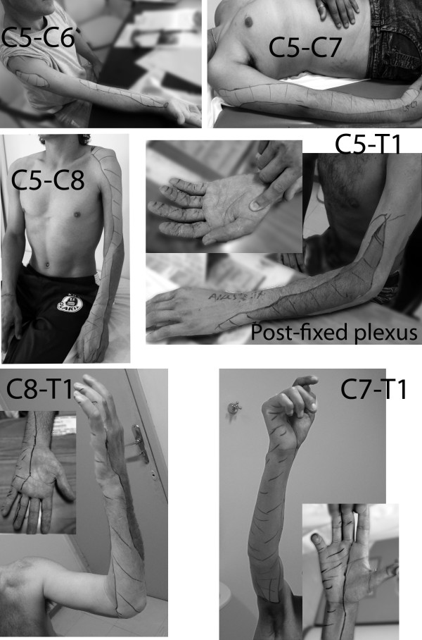
We review our experience treating 335 adult patients with supraclavicular brachial plexus injuries over a 7-year period at the University of Southern Santa Catarina, in Brazil. Patients were categorized into 8 groups, according to functional deficits and roots injured: C5-C6, C5-C7, C5-C8 (T1 Hand), C5-T1 (T2 Hand), C8-T1, C7-T1, C6-T1, and total palsy. To restore function, nerve grafts, nerve transfers, and tendon and muscle transfers were employed. Patients with either upper- or lower-type partial injuries experienced considerable functional return. In total palsies, if a root was available for grafting, 90% of patients had elbow flexion restored, whereas this rate dropped to 50% if no roots were grafted and only nerve transfers performed. Pain resolution should be the first priority, and root exploration and grafting helped to decrease or eliminate pain complaints within a short time of surgery.


Figures

Figure 1
Figure 1
Zones of lost protective sensation with the different types of brachial plexus palsy. Mapping was performed following assessments using Semmes-Weinstein monofilaments. In those with a C5-C6 injury, hand sensation was totally preserved. In the C5-C7 injury group, hand sensation was decreased, but still was within the normal range. A longitudinal area of absent protective sensation was present on the lateral aspect of the forearm and arm. In the C5-C8 palsy group, there was a similar longitudinal area along the lateral side of the arm and forearm, associated with no protective sensation. The dorsal side of the hand also was now markedly affected. On the palmar aspect of the hand, sensation decreased to a variable degree. Almost half of the patients had normal sensation, and the remaining half experienced thumb anesthesia. In those with a C5-T1 lesion with post-fixation of the plexus, only a small zone was observed in which there was preserved sensation over the medial side of the forearm. Hand sensation was markedly reduced. The thumb was anesthetized, but protective sensation was demonstrated in the long fingers (inset). In the C8-T1 palsy group, a loss of protective sensation was evident on the medial side of the forearm and in the ulnar fingers. In the C7-T1 injury group, the inner aspect of the arm also was affected, together with additional involvement of the long finger.
Figure 2
Figure 2
Intra-operative view of a left axillary approach to neurotize the anterior division (AD) of the axillary nerve and the teres minor motor branch. Through this same approach, ulnar nerve fascicles are concomitantly transferred to the biceps motor branch. (PD) posterior division of the axillary nerve and its branches: (TM) teres minor motor branch, (POD) branch to the posterior deltoid muscle, and (CB), the upper arm lateral cutaneous nerve.
Figure 3
Figure 3
A) Intra-operative view of transferring the pronator quadratus motor branch (i.e. anterior interosseous nerve) to the extensor carpi radialis motor branch (ECRB). B) After sectioning, the proximal stump of the anterior interosseous nerve (AIN) was flipped proximally for suturing to the extensor carpi radialis brevis motor branch, which was dissected and sectioned proximally, and flipped distally. The distal stump of the anterior interosseous nerve was sutured to a motor fascicle of the median nerve, (MN) end-to-end, to restore pronation.
Figure 4
Figure 4
Intra-operative view of a pulley dermodesis for correction of metacarpophalangeal hyperextension in a patient with a lower type palsy of the right brachial plexus. After resection of a cutaneous ellipse centered on the distal palmar crease, the A1 pulley was sutured to the palmar aponeurosis and proximal dermis.
Figure 5
Figure 5
Schematic representation of procedures to restore sensation on the ulnar side of the hand in patients with a lower-type palsy of the brachial plexus. Either the palmar cutaneous branch of the median nerve was transferred to the dorsal branch of the ulnar nerve, or the proper digital nerve of the little finger was sutured to fascicles of the median nerve to the palm, either in association or not in association with fascicles raised from the proper ulnar digital nerve of the index finger.

References

    1. Robotti E, Longhi P, Verna G, Bocchiotti G. Brachial plexus surgery: an historical perspective. Hand Clin. 1995;6:517–533. - PubMed
    1. Bertelli JA, Ghizoni MF. Use of clinical signs and computed tomography myelography findings in detecting and excluding nerve root avulsion in complete brachial plexus palsy. J Neurosurg. 2006;6:835–842. doi: 10.3171/jns.2006.105.6.835. - DOI - PubMed
    1. Bertelli JA, Ghizoni MF. Contralateral motor rootlets and ipsilateral nerve transfers in brachial plexus reconstruction. J Neurosurg. 2004;6:770–778. doi: 10.3171/jns.2004.101.5.0770. - DOI - PubMed
    1. Bertelli JA, Ghizoni MF. Reconstruction of C5 and C6 brachial plexus avulsion injury by multiple nerve transfers: spinal accessory to suprascapular, ulnar fascicles to biceps branch, and triceps long or lateral head branch to axillary nerve. J Hand Surg Am. 2004;6:131–139. doi: 10.1016/j.jhsa.2003.10.013. - DOI - PubMed
    1. Bertelli JA, Ghizoni MF. Nerve root grafting and distal nerve transfers for C5-C6 brachial plexus injuries. J Hand Surg Am. 2010;6:769–75. doi: 10.1016/j.jhsa.2010.01.004. - DOI - PubMed