

RTOG sarcoma radiation oncologists reach consensus on gross tumor volume and clinical target volume on computed tomographic images for preoperative radiotherapy of primary soft tissue sarcoma of extremity in Radiation Therapy Oncology Group studies
- PMID: 21676552
- PMCID: PMC3205346
- DOI: 10.1016/j.ijrobp.2011.04.038
RTOG sarcoma radiation oncologists reach consensus on gross tumor volume and clinical target volume on computed tomographic images for preoperative radiotherapy of primary soft tissue sarcoma of extremity in Radiation Therapy Oncology Group studies
Abstract
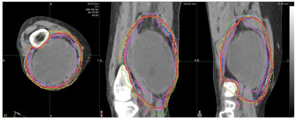
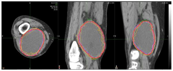
Objective: To develop a Radiation Therapy Oncology Group (RTOG) atlas delineating gross tumor volume (GTV) and clinical target volume (CTV) to be used for preoperative radiotherapy of primary extremity soft tissue sarcoma (STS).
Methods and materials: A consensus meeting was held during the RTOG meeting in January 2010 to reach agreement about GTV and CTV delineation on computed tomography (CT) images for preoperative radiotherapy of high-grade large extremity STS. Data were presented to address the local extension of STS. Extensive discussion ensued to develop optimal criteria for GTV and CTV delineation on CT images.
Results: A consensus was reached on appropriate CT-based GTV and CTV. The GTV is gross tumor defined by T1 contrast-enhanced magnetic resonance images. Fusion of magnetic resonance and images is recommended to delineate the GTV. The CTV for high-grade large STS typically includes the GTV plus 3-cm margins in the longitudinal directions. If this causes the field to extend beyond the compartment, the field can be shortened to include the end of a compartment. The radial margin from the lesion should be 1.5 cm, including any portion of the tumor not confined by an intact fascial barrier, bone, or skin surface.
Conclusion: The consensus on GTV and CTV for preoperative radiotherapy of high-grade large extremity STS is available as web-based images and in a descriptive format through the RTOG. This is expected to improve target volume consistency and allow for rigorous evaluation of the benefits and risks of such treatment.
Copyright © 2011 Elsevier Inc. All rights reserved.
Figures
Comment in
-
In regard to RTOG sarcoma radiation oncologists reach consensus on gross tumor volume and clinical target volume on computed tomographic images for preoperative radiotherapy of primary soft tissue sarcoma of extremity in Radiation Therapy Oncology Group studies: in regard to Wang et al (Int J Radiat Oncol Biol Phys 2011;81:e525-e528).Int J Radiat Oncol Biol Phys. 2012 Jun 1;83(2):483. doi: 10.1016/j.ijrobp.2012.01.059. Int J Radiat Oncol Biol Phys. 2012. PMID: 22579375 No abstract available.
References
-
- Mackie TR, Kapatoes J, Ruchala K, et al. Image guidance for precise conformal radiotherapy. Int J Radiat Oncol Biol Phys. 2003;56:89–105. - PubMed
-
- Yan D, Lockman D, Martinez A, et al. Computed tomography guided management of interfractional patient variation. Semin Radiat Oncol. 2005;15:168–79. - PubMed
-
- Mackie TR, Balog J, Ruchala K, et al. Tomotherapy. Semin Radiat Oncol. 1999;9:108–17. - PubMed
-
- Mohan R, Zhang X, Wang H, et al. Use of deformed intensity distributions for on-line modification of image-guided IMRT to account for interfractional anatomic changes. Int J Radiat Oncol Biol Phys. 2005;61:1258–66. - PubMed
-
- Jaffray DA. Emergent technologies for 3-dimensional image-guided radiation delivery. Semin Radiat Oncol. 2005;15:208–16. - PubMed
Publication types
MeSH terms
Substances
Grants and funding
LinkOut - more resources
Full Text Sources
Other Literature Sources
Medical
