. 2011 Oct;25(10):3416-25.
doi: 10.1096/fj.11-186346. Epub 2011 Jun 15.

The SUMO E3-ligase PIAS1 couples reactive oxygen species-dependent JNK activation to oxidative cell death

Affiliations

The SUMO E3-ligase PIAS1 couples reactive oxygen species-dependent JNK activation to oxidative cell death

Beatriz B Leitao et al. FASEB J. 2011 Oct.

Abstract

Human endometrial stromal cells (HESCs) exposed to reactive oxygen species (ROS) mount a hypersumoylation response in a c-Jun N-terminal kinase (JNK)-dependent manner. The mechanism that couples JNK signaling to the small ubiquitin-related modifier (SUMO) pathway and its functional consequences are not understood. We show that ROS-dependent JNK activation converges on the SUMO pathway via PIAS1 (protein inhibitor of activated STAT1). Unexpectedly, PIAS1 knockdown not only prevented ROS-dependent hypersumoylation but also enhanced JNK signaling in HESCs. Conversely, PIAS overexpression increased sumoylation of various substrates, including c-Jun, yet inhibited basal and ROS-dependent JNK activity independently of its SUMO ligase function. Expression profiling demonstrated that PIAS1 knockdown enhances and profoundly modifies the transcriptional response to oxidative stress signals. Using a cutoff of 2-fold change or more, a total of 250 ROS-sensitive genes were identified, 97 of which were not dependent on PIAS1. PIAS1 knockdown abolished the regulation of 43 genes but also sensitized 110 other genes to ROS. Importantly, PIAS1 silencing was obligatory for the induction of several cellular defense genes in response to oxidative stress. In agreement, PIAS1 knockdown attenuated ROS-dependent caspase-3/7 activation and subsequent apoptosis. Thus, PIAS1 determines the level of JNK activity in HESCs, couples ROS signaling to the SUMO pathway, and promotes oxidative cell death.


Figures

Figure 1.
Figure 1.
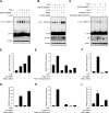
ROS-dependent JNK1 activation correlates with cellular hypersumoylation and PIAS1 modification. A) Primary HESC cultures, undifferentiated or decidualized with 8-br-cAMP and MPA for 1, 2, and 4 d, were exposed to 250 μM H2O2 for 30 min. Whole-cell extracts were separated by SDS–PAGE and probed by Western blotting with antibodies against SUMO-1, PIAS1, total JNK, and phosphorylated JNK (p-JNK). β-Actin served as a loading control. B) Endogenous PIAS1 immunoprecipitated from HESCs exposed to 250 μM H2O2 for 30 min was incubated with or without λ PPase (200 U/μl, 30 min, 30°C), separated on SDS-PAGE, and probed for PIAS1. Expression levels of PIAS1 in the inputs were verified by Western blot analysis. C) Primary HESC cultures were transfected with HA-tagged PIAS1 either with NT or JNK-targeting siRNA and exposed to 250 μM H2O2 for 30 min. Whole-cell extracts were subjected to Western blot analysis with antibodies against HA, p-JNK, and β-actin. D) HA-PIAS1 immunoprecipitated from COS-1 cells, first cotransfected with or without an expression vector coding for ΔMEKK:ER*-myc and then treated with 100 nM 4-hydroxytamoxifen (4-OHT) for 30 min, was incubated with or without λ PPase (200 U/μl, 30 min, 30°C), separated on SDS-PAGE. and probed with an anti-HA antibody. Expression levels of PIAS1 in the inputs were verified by Western blot analysis. E) JNK phosphorylates PIAS1 in vitro. HA-PIAS1 immunopurified from transfected COS-1 cells was subjected to an in vitro kinase assay with recombinant JNK. Immunoprecipitation of PIAS1 was verified by Western blot analysis of the HA tag.
Figure 2.
Figure 2.
PIAS1 mediates the ROS-dependent cellular hypersumoylation response and is a negative regulator of JNK. A) Primary HESC cultures, transfected either with NT or PIAS1-targeting siRNA, were exposed to 250 μM H2O2 for 10 or 30 min. Whole-cell protein extracts were subjected to Western blot analysis with antibodies against SUMO-1, PIAS1, phosphorylated JNK (p-JNK), and total JNK. β-Actin served as loading control. B) Primary HESC cultures transfected with a noncoding vector or Flag-tagged PIAS1 were exposed to 250 μM H2O2 for 10 or 30 min, and total cell lysates were immunoprobed for SUMO-1, Flag tag, p-JNK, and total JNK. C) Primary HESC cultures were transfected with an expression vector coding for Δ MEKK:ER*-myc either with or without HA-PIAS1 and treated with 100 nM 4-OHT for 30 min. PIAS1 expression and JNK phosphorylation were assessed by Western blot analysis. D) Primary cultures, transfected with a noncoding vector or with an expression vector encoding either WT PIAS1 or ligase-deficient (W372A/C351S) mutant, were pulsed with 250 μM H2O2 for 30 min. JNK phosphorylation relative to total JNK levels was measured with PathScan phospho- and total SAPK/JNK Chemiluminescent Sandwich ELISA kits, respectively. Activity is measured in relative light units (RLU) and normalized to the levels in untreated cells transfected with a noncoding vector. Data represent means ± sd of triplicate determinations from 3 independent experiments.
Figure 3.
Figure 3.
PIAS1 modulates c-Jun sumoylation and activity. A) Primary HESCs, cotransfected with expressing vectors encoding EGFP-SUMO-1 and c-Jun, were exposed to 250 μM H2O2 for the indicated time periods, and whole-cell lysates were immunoprobed for c-Jun and β-actin. B) Primary cultures were transfected with expressing vectors coding for EGFP-SUMO-1 and c-Jun in combination with either pSG5-PIAS1 or PIAS1 siRNA. Cells were then treated with 250 μM H2O2 for 30 min; total cell lysates were immunoprobed for c-Jun, PIAS1, and β-actin. C) HESCs were transfected with expression vectors encoding EGFP-SUMO-1 and c-Jun in combination with plasmids encoding either HA-tagged WT PIAS1 or ligase mutant (HA-PIAS1 W372A/C351S). Cells were exposed to 250 μM H2O2 for 30 min and subjected to Western blot analysis. D–I) PIAS1 modulates the expression of JUN and RRAD in response to ROS-dependent JNK activation. Primary HESCs were transfected either with PIAS1 siRNA (D, G), expression vector encoding WT or ligase-deficient PIAS1 (E, H), or siRNA targeting JNK (F, I). After 2 d, cultures were treated with 250 μM H2O2 for 8 h, and the abundance of JUN (D–F) and RADD (G–I) transcripts was determined by RTQ-PCR. Data are presented as mean ± sd fold induction. Results are representative of 3 independent experiments.
Figure 4.
Figure 4.
PIAS1 modulates the transcriptional response to ROS in HESCs. A) Primary HESC cultures transfected with either NT or PIAS1-targeting siRNA were treated with 250 μM H2O2 for 8 h, and total RNA was subjected to genome-wide expression profiling. Venn diagrams indicate number of significantly up- and down-regulated ROS-responsive genes dependent or independent of PIAS1 knockdown. Left and right panels indicate the number of genes regulated ≥2- or ≥1.2-fold, respectively. B) For validation, transcript levels of selective ROS-sensitive genes were determined by RTQ-PCR in HESC cultures first transfected with either NT or PIAS1-targeting siRNA and then treated with 250 μM H2O2 for the indicated time periods. Data are presented as mean ± sd fold induction; results are representative of 3 biological repeats.
Figure 5.
Figure 5.
PIAS1 promotes oxidative cell death. A) Percentage of apoptotic cells containing sub-G1 DNA was determined by flow cytometry analysis of primary cultures first transfected with NT or PIAS1-targeting siRNA and then treated with 250 μM H2O2 for 24 h. Results are means ± sd of 3 independent experiments. B) Caspase 3 and 7 activity in response to H2O2 treatment was determined by ELISA in HESC cultures first transfected with NT or PIAS1-targeting siRNA. C) Primary cultures, transfected with a noncoding expression vector or a plasmid encoding the SENP2 isopeptidase, were treated with 250 μM H2O2 for 4 h; activated caspase-3 and caspase-7 levels were determined by ELISA.
Figure 6.
Figure 6.
Schematic diagram summarizing the bidirectional role of PIAS1 in regulating JNK-dependent oxidative stress responses. PIAS1 is a target as well as a negative regulator of the activated JNK pathway in HESCs exposed to ROS. PIAS1 also drives the associated hypersumoylation response and inhibits the expression of defense genes, and thereby promotes oxidative apoptosis in HESCs.

References

    1. Cheng J., Perkins N. D., Yeh E. T. (2005) Differential regulation of c-Jun-dependent transcription by SUMO-specific proteases. J. Biol. Chem. 280, 14492–14498 - PubMed
    1. Gellersen B., Brosens I. A., Brosens J. J. (2007) Decidualization of the human endometrium: mechanisms, functions, and clinical perspectives. Semin. Reprod. Med. 25, 445–453 - PubMed
    1. Brosens J. J., Gellersen B. (2006) Death or survival–progesterone-dependent cell fate decisions in the human endometrial stromal. J. Mol. Endocrinol. 36, 389–398 - PubMed
    1. Gellersen B., Brosens J. (2003) Cyclic AMP and progesterone receptor cross-talk in human endometrium: a decidualizing affair. J. Endocrinol. 178, 357–372 - PubMed
    1. Brosens J. J., Parker M. G., McEnroe A., Pijnenborg R., Brosens I. A. (2009) A role for menstruation in preconditioning the uterus for successful pregnancy. Am. J. Obstet. Gynecol. 200, 615.e1–615.e6 - PubMed

Publication types

MeSH terms

Substances