. 2011 Jul 15;187(2):1023-30.
doi: 10.4049/jimmunol.1004195. Epub 2011 Jun 15.

Anticardiac myosin immunity and chronic allograft vasculopathy in heart transplant recipients

Affiliations

Anticardiac myosin immunity and chronic allograft vasculopathy in heart transplant recipients

Safa Kalache et al. J Immunol. .

Abstract

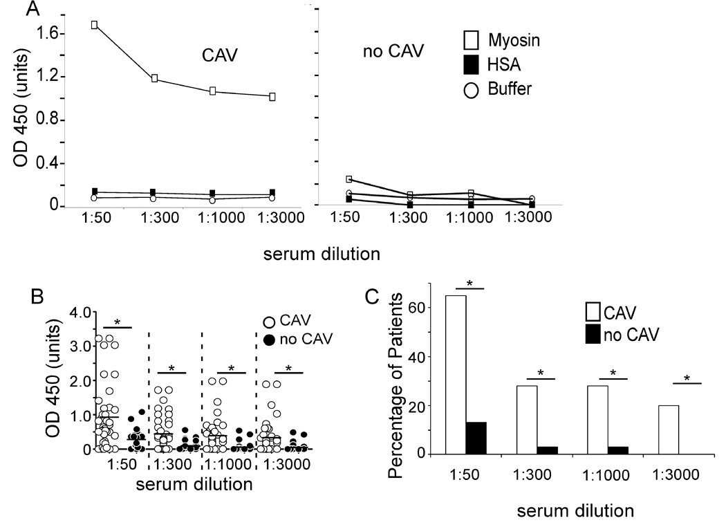
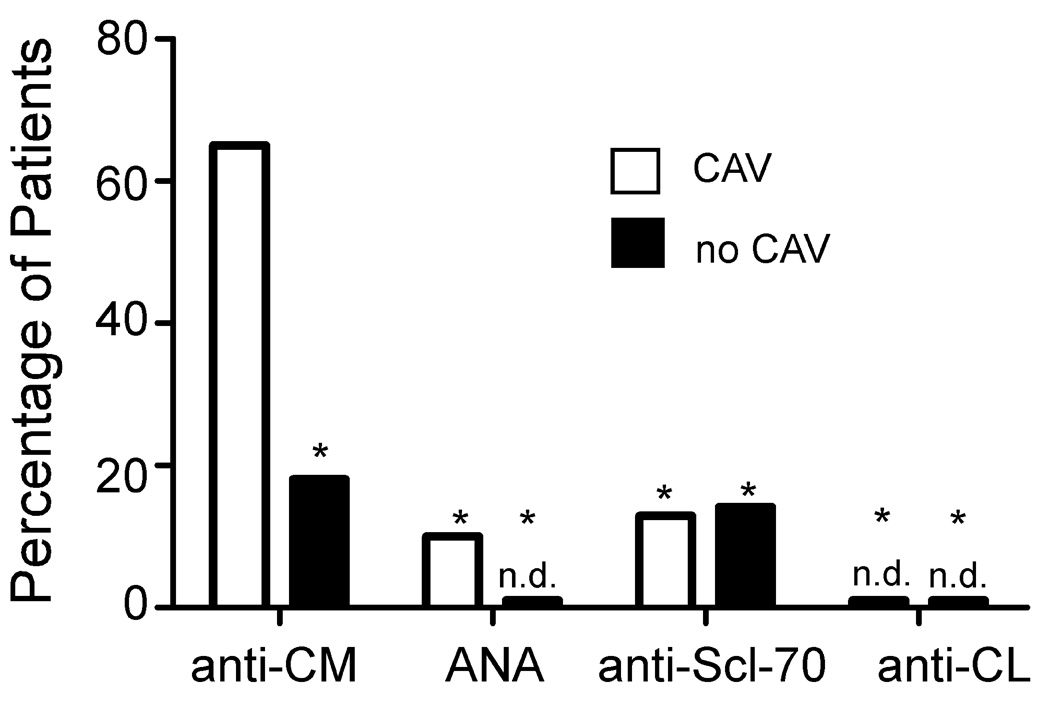
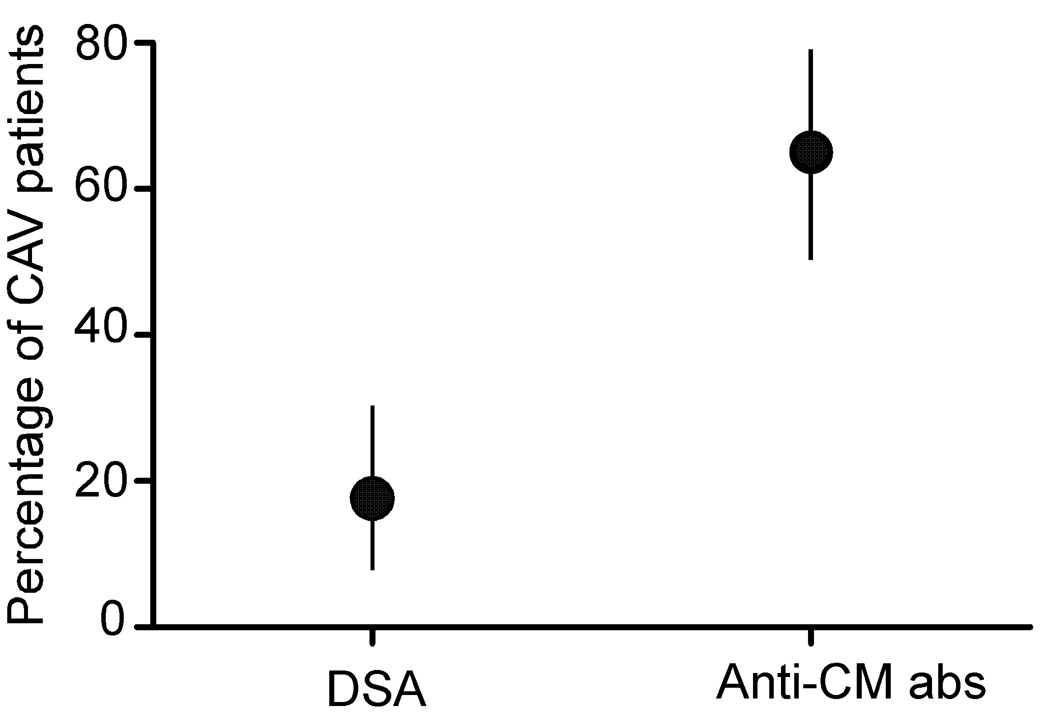
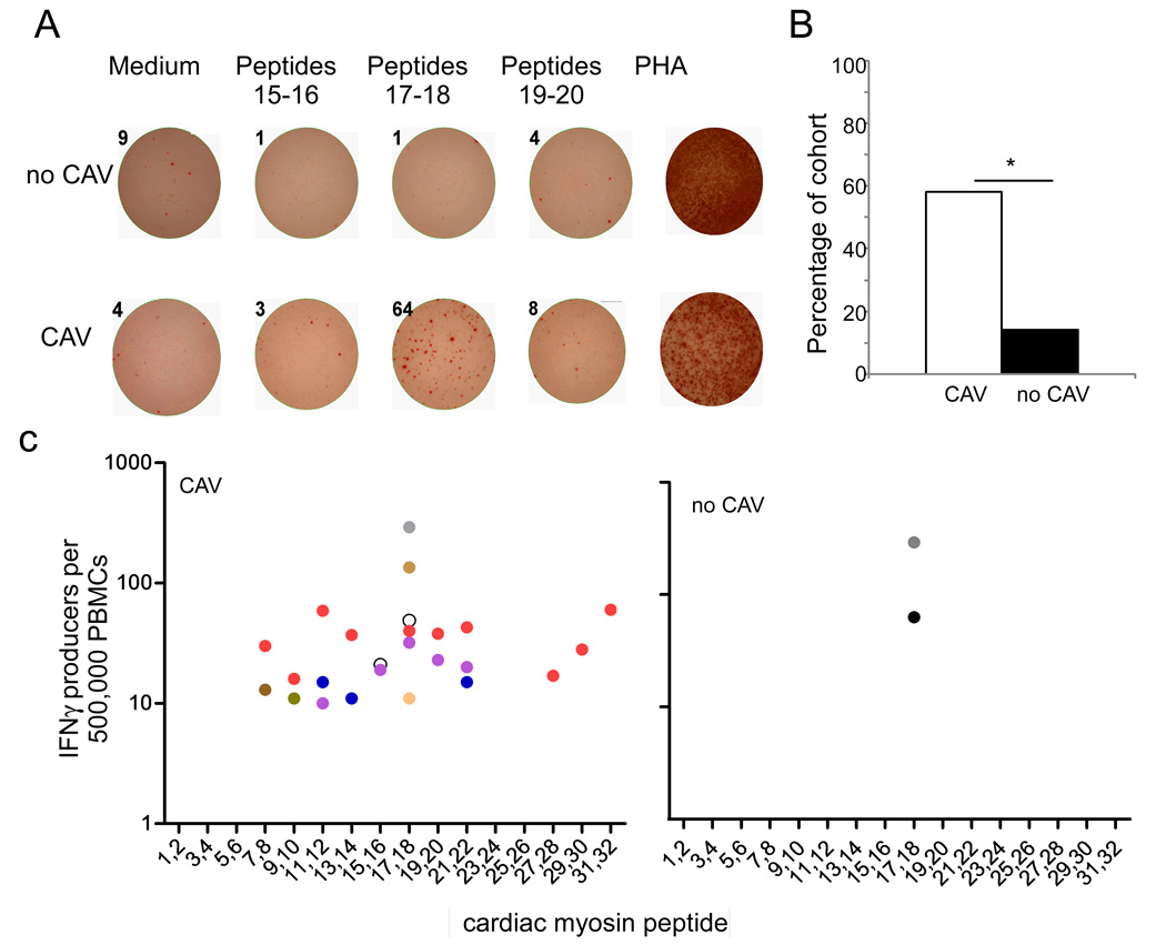
Chronic allograft vasculopathy (CAV) contributes to heart transplant failure, yet its pathogenesis is incompletely understood. Although cellular and humoral alloimmunity are accepted pathogenic mediators, animal models suggest that T cells and Abs reactive to graft-expressed autoantigens, including cardiac myosin (CM), could participate. To test the relationship between CAV and anti-CM autoimmunity in humans, we performed a cross-sectional study of 72 heart transplant recipients: 40 with CAV and 32 without. Sera from 65% of patients with CAV contained anti-CM Abs, whereas <10% contained Abs to other autoantigens (p < 0.05), and only 18% contained anti-HLA Abs (p < 0.05 versus anti-CM). In contrast, 13% of sera from patients without CAV contained anti-CM Abs (p < 0.05; odds ratio [OR], associating CAV with anti-CM Ab = 13, 95% confidence interval [CI] 3.79-44.6). Multivariable analysis confirmed the association to be independent of time posttransplant and the presence of anti-HLA Abs (OR = 28, 95% CI 5.77-133.56). PBMCs from patients with CAV responded more frequently to, and to a broader array of, CM-derived peptides than those without CAV (p = 0.01). Detection of either CM-peptide-reactive T cells or anti-CM Abs was highly and independently indicative of CAV (OR = 45, 95% CI 4.04-500.69). Our data suggest detection of anti-CM immunity could be used as a biomarker for outcome in heart transplantation recipients and support the need for further studies to assess whether anti-CM immunity is a pathogenic mediator of CAV.


Figures

Figure 1
Figure 1. Anti-CM antibodies are found in serum from patients with CAV
Sera from 40 patients with CAV and 32 patients without CAV were tested for anti- CM antibodies at serum dilutions of 1:50, 1:300, 1:1000, and 1:3000. A. Representative ELISA results of serum from one patient with CAV (left) and one patient without CAV (right). Open squares represent reactivity to CM, solid squares HSA and open circles buffer alone. B. ELISA results from all CAV and nCAV patients at each serum dilution (1:50, 1:300, 1:1000, 1:3000). C. Summary depicting the percentage of CAV and nCAV patients with anti-CM immunity of OD>0.5 (*p<0.05).
Figure 2
Figure 2. Anti-CM antibodies are more prevalent than antibodies to other common autoantigens in heart transplant recipients
Results for anti-CM antibodies (CAV, n=40; no CAV, n=32, dilution 1:50) in Figure 1 are re-plotted along with results of assays performed on the same sera testing for reactivity to ANA, anti-Scl-70, and anti-cardiolipin (CL) antibodies by ELISA. Sera from patients with CAV more commonly contained anti-CM antibodies than any of the other antibodies tested (*p <0.05). n.d., none detected.
Figure 3
Figure 3. Anti-CM antibodies are not associated with specific recipient HLA alleles or the number of HLA mismatches between donor and recipient
The percentages of anti-CM antibody positive (+) and anti-CM antibody negative (−) patients expressing each HLA-A (A), HLA-B (B), and HLA-DR (C) allele are shown. Anti-CM antibodies did not associate with any HLA allele in the cohort. Panel D depicts the number of HLA mismatches between donor and recipient at A, B, and DR loci (0–6) plotted against the presence or absence of anti-CM antibody in the serum. The mean number of mismatches is not different between those with and without anti-CM antibodies (vertical line, p=ns).
Figure 4
Figure 4. Anti-HLA antibodies are equally prevalent in those with and without CAV
Sera from 40 patients with CAV and 32 patients without CAV were screened for the presence of anti-HLA antibodies using luminex. There was no statistically significant difference in percentage of patients who screened positive for class I alone, class II alone, class I and class II, or either class I or II between the two groups. p=ns for all groups.
Figure 5
Figure 5. Higher prevalence of anti-CM antibodies than Donor Specific Antibodies (DSA) in patients with CAV
Graphical depiction of the prevalence of donor-HLA reactive antibodies and anti-CM antibodies, with 95% confidence intervals (vertical lines), in the 20 CAV patients that screened positive for anti-HLA antibodies by screening tests. Because the confidence intervals do not overlap the differences are statistically significant.
Figure 6
Figure 6. Stronger T cell reactivity to CM in patients with CAV
A. Representative IFNγ ELISPOT wells in which PBMCs obtained from one patient without CAV (no CAV, top row) and one with CAV (bottom row) were stimulated with medium alone or 3 different pairs of CM-derived peptides. PHA served as a positive control. Numbers above each well are the spots detected. B. The prevalence of T cell reactivity to CM as defined by >10 IFNγ producers per 500,000 PBMCs in response to at least one peptide is shown for 18 patients with CAV and 13 patients without CAV (*p<0.05). C. Peptide mapping of PBMCs from CAV patients (left) and nCAV patients (right). Each color represents the responses of a different patient.
Figure 7
Figure 7. Treg mediated suppression does explain the absence of CM-peptide reactive T cell immunity
A. Representative flow cytometry plots of PBMC before (above) and after (below) removal of CD25+ cells using magnetic beads, verifying depletion of the CD4+ CD25+ Foxp3+ subset (right panels). B. IFNγ ELISPOT assays for reactivity to CM peptides 17–18 using pre-depletion and post-depletion PBMC from six different patients, 4 without detectable responses pre-depletion (left of dotted line) and 2 with detectable responses pre-depletion (right of dotted line). PBMC from an additional 6 patients without detectable responses pre-depletion showed identical results (no new responses after depletion, not shown).

References

    1. http://optn.transplant.hrsa.gov.
    1. Johnston DR, Sayegh MH, Madsen JC. Overcoming cardiac allograft vasculopathy (CAV) by inducing tolerance. Front Biosci. 2002;7:e116–e118. - PubMed
    1. Mehra MR. Contemporary concepts in prevention and treatment of cardiac allograft vasculopathy. Am J Transplant. 2006;6:1248–1256. - PubMed
    1. Chen Y, Demir Y, Valujskikh A, Heeger PS. The male minor transplantation antigen preferentially activates recipient CD4+ T cells through the indirect presentation pathway in vivo. J Immunol. 2003;171:6510–6518. - PubMed
    1. Sahara H, Shoji T, Ng CY, Weiss MJ, Muniappan A, Guenther DA, Houser SL, Pujara AC, Sayre JK, Wain JC, Sachs DH, Madsen JC, Allan JS. The role of indirect recognition of MHC class I and II allopeptides in a fully mismatched miniature swine model of lung transplantation. Transplant Proc. 2006;38:3256–3258. - PMC - PubMed

Publication types

MeSH terms