. 2011 Jun 15;31(24):8708-12.
doi: 10.1523/JNEUROSCI.6565-10.2011.

Cerebellar-dependent learning in larval zebrafish

Affiliations

Cerebellar-dependent learning in larval zebrafish

Mark Aizenberg et al. J Neurosci. .

Abstract

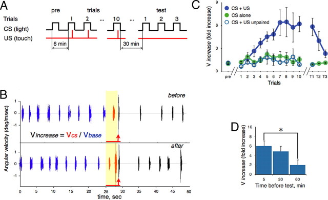
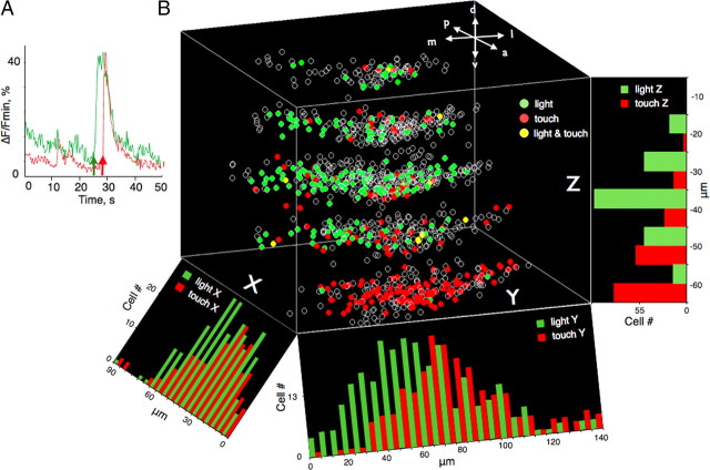
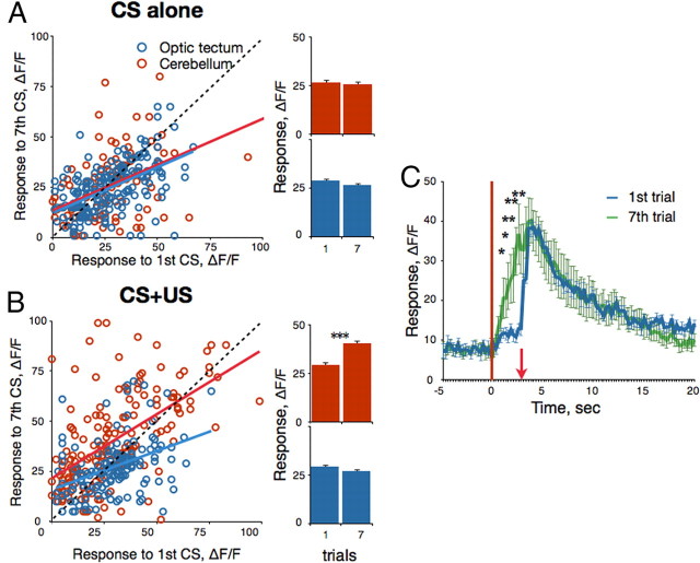
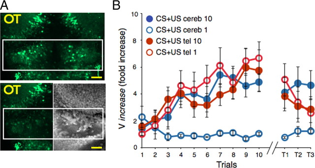
Understanding how neuronal network activity contributes to memory formation is challenged by the complexity of most brain circuits and the restricted ability to monitor the activity of neuronal populations in vivo. The developing zebrafish (Danio rerio) is an animal model that circumvents these problems, because zebrafish larvae possess a rich behavioral repertoire and an accessible brain. Here, we developed a classical conditioning paradigm in which 6- to 8-d-old larvae develop an enhanced motor response to a visual stimulus (conditioned stimulus, CS) when it is paired with touch (unconditioned stimulus, US). Using in vivo calcium imaging we demonstrate that CS and US activate different subsets of neurons in the cerebellum; their activity, modulated by learning two-photon laser ablation, revealed that the cerebellum is involved in acquisition and extinction, but not the retention, of this memory.


Figures

Figure 1.
Figure 1.
Classical conditioning in larval zebrafish. A, Conditioning protocol. In the first trial (pre) a single CS was presented to examine the animal's response to the CS alone. Ten paired CS+US trials were presented with a 6 min intertrial interval (ITI). Three test trials (CS alone) were presented after various delays. B, Sample analysis of conditioned and unconditioned responses (CR and UR, respectively). The tail angular velocity before (upper trace) and after (lower trace) learning was compared. The UR can be seen after US presentation (red arrows). The red line represents the CS. C, The effect of paired (CS+US), unpaired (CS+US unpaired), and CS alone trials on the CR. Each point represents the mean Vincrease value for all fish in a given trial ± SEM. Conditioning resulted in a significantly enhanced CR that grew in magnitude over the 10 trials and was rapidly extinguished by three test trials. Training with CS alone or unpaired CS+US did not significantly alter the CR. D, Memory decline over time. Fish were conditioned, and each group was tested twice (ITI = 4 min) either 5, 30, or 60 min after the last trial. Each bar represents mean Vincrease value for two trials ± SEM. *p < 0.05.
Figure 2.
Figure 2.
A, Change in fluorescence averaged across 25 light- and touch-evoked cerebellar neurons (green and red lines, respectively) as a function of time. The green arrow represents the onset of the visual stimulus (CS). The red arrow depicts the timing of touch delivery. B, Three-dimensional functional map of the left cerebellar hemisphere. The zero point on the z-axis is located at the dorsal surface of the cerebellum. Each z-section is separated by 10 μm. The intersection between x- and y-axes is the point where the midline between the hemispheres crosses the border to the optic tectum. Each circle depicts the position of an individual neuron. Green circles represent the light-driven neurons. Red circles represent touch-evoked neurons. Yellow circles represent neurons that responded to both visual and tactile stimuli. Orientation is as indicated: a, anterior; p, posterior; m, medial; l, lateral; d, dorsal, v, ventral. Histograms depict the density of light- and touch-evoked neurons (green and red bars, respectively) projected on x-, y-, and z-axes. Note that the density of touch-driven cells gradually increases in the dorsolateral direction, whereas light-evoked neurons are most densely packed at a depth of 40 μm.
Figure 3.
Figure 3.
Learning-dependent changes in visually driven activity. A, Neurons in the cerebellum (red) and optic tectum (blue) display relatively stable visual responses to a repetitively presented CS. B, Cerebellar, but not tectal, neurons, increase their responses to the CS during conditioning (CS+US). In A and B, each dot represents the response of an individual neuron (peak height) to the CS in trial 7 as a function of the response in trial 1. Color lines are linear trend lines. Bars on the right depict averaged responses in trials 1 and 7. ΔF/Fmin, percentage ± SEM; ***p < 0.001. C, Some neurons responded to the US only at the beginning of the training session but developed an additional peak of activity in response to CS after conditioning. An average response of 12 neurons from six fish (ΔF/Fmin, percentage ± SEM) in the first (blue) and seventh (green) trials is shown. Onsets of the CS and US are represented by the vertical red line and red arrow, respectively. *p < 0.05; **p < 0.01.
Figure 4.
Figure 4.
Requirement of the cerebellum, but not the telencephalon, for learning acquisition. A, Two-photon ablation of the telencephalon (left) and cerebellum (right). Top is a z-projection of confocal sections taken at depths of 0–40 μm. Bottom shows the left side of the same fish after ablation and the right side of a fixed brain stained with SYTO 14 (white rectangle shows the ablated region). Scale bar, 20 μm. B, Graph plots the CR (mean Vincrease ± SEM) as a function of trials. Bilateral ablation of the telencephalon performed immediately after the first (open red circles) or 10th (filled red circles) trial did not significantly impair acquisition or retention of memory. Bilateral lesions in the cerebellum after the first trial (open blue circles) blocked acquisition of the CR. Ablation after the 10th trial (filled blue circles) did not affect memory retention but impaired extinction during the three test trials.

References

    1. Asakawa K, Suster ML, Mizusawa K, Nagayoshi S, Kotani T, Urasaki A, Kishimoto Y, Hibi M, Kawakami K. Genetic dissection of neural circuits by Tol2 transposon-mediated Gal4 gene and enhancer trapping in zebrafish. Proc Natl Acad Sci U S A. 2008;105:1255–1260. - PMC - PubMed
    1. Bae YK, Kani S, Shimizu T, Tanabe K, Nojima H, Kimura Y, Higashijima S, Hibi M. Anatomy of zebrafish cerebellum and screen for mutations affecting its development. Dev Biol. 2009;330:406–426. - PubMed
    1. Baier H, Scott EK. Genetic and optical targeting of neural circuits and behavior — zebrafish in the spotlight. Curr Opin Neurobiol. 2009;19:553–560. - PMC - PubMed
    1. Burgess HA, Granato M. Sensorimotor gating in larval zebrafish. J Neurosci. 2007;27:4984–4994. - PMC - PubMed
    1. Darland T, Dowling JE. Behavioral screening for cocaine sensitivity in mutagenized zebrafish. Proc Natl Acad Sci U S A. 2001;98:11691–11696. - PMC - PubMed

Publication types

Substances

LinkOut - more resources