. 2011 Jun 15;31(24):8998-9009.
doi: 10.1523/JNEUROSCI.6684-10.2011.

Essential roles of enteric neuronal serotonin in gastrointestinal motility and the development/survival of enteric dopaminergic neurons

Affiliations

Essential roles of enteric neuronal serotonin in gastrointestinal motility and the development/survival of enteric dopaminergic neurons

Zhishan Li et al. J Neurosci. .

Abstract

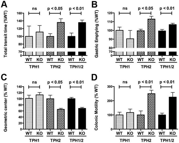
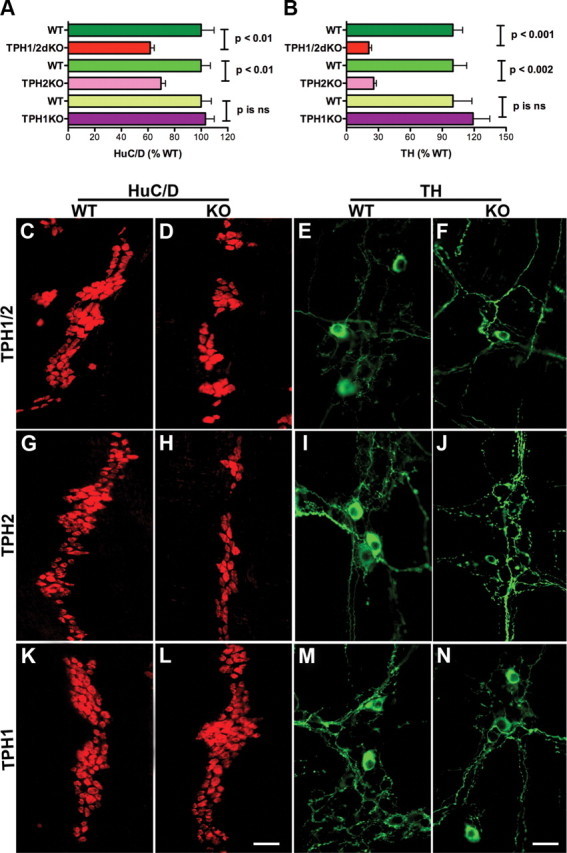
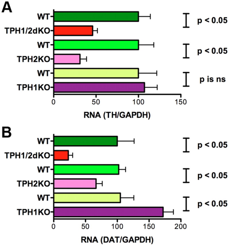
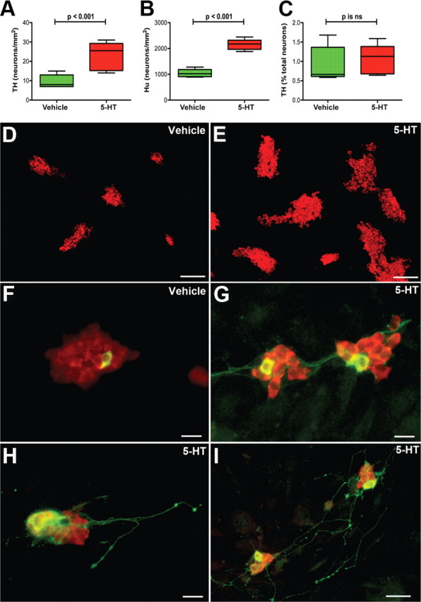
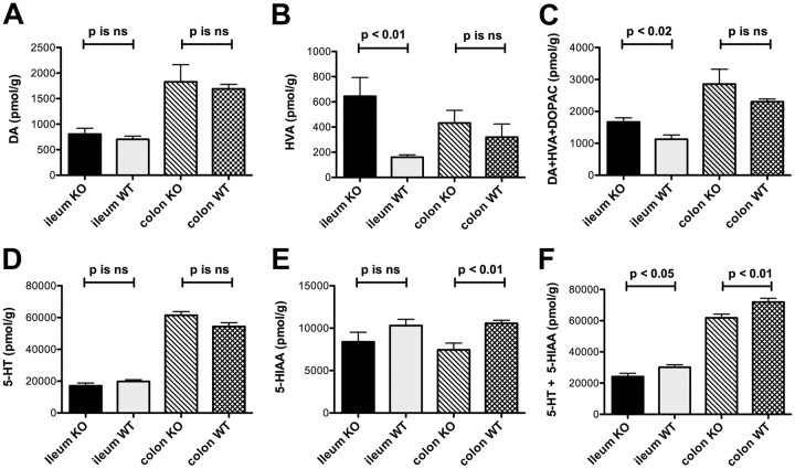
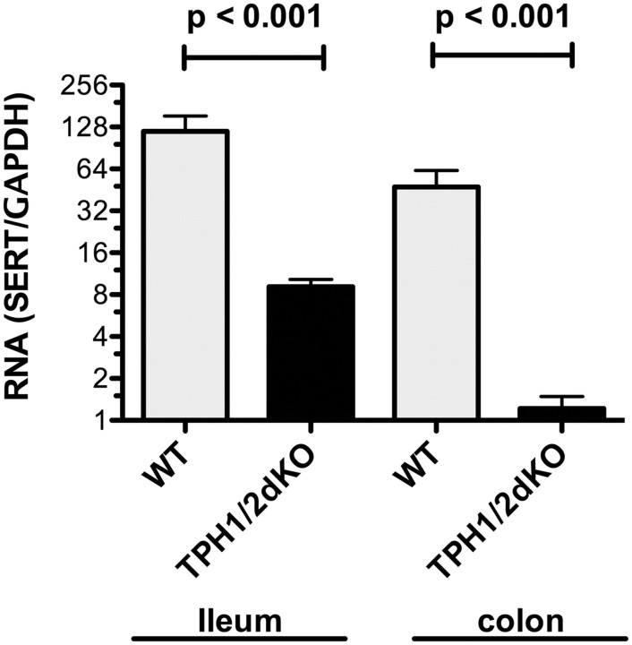
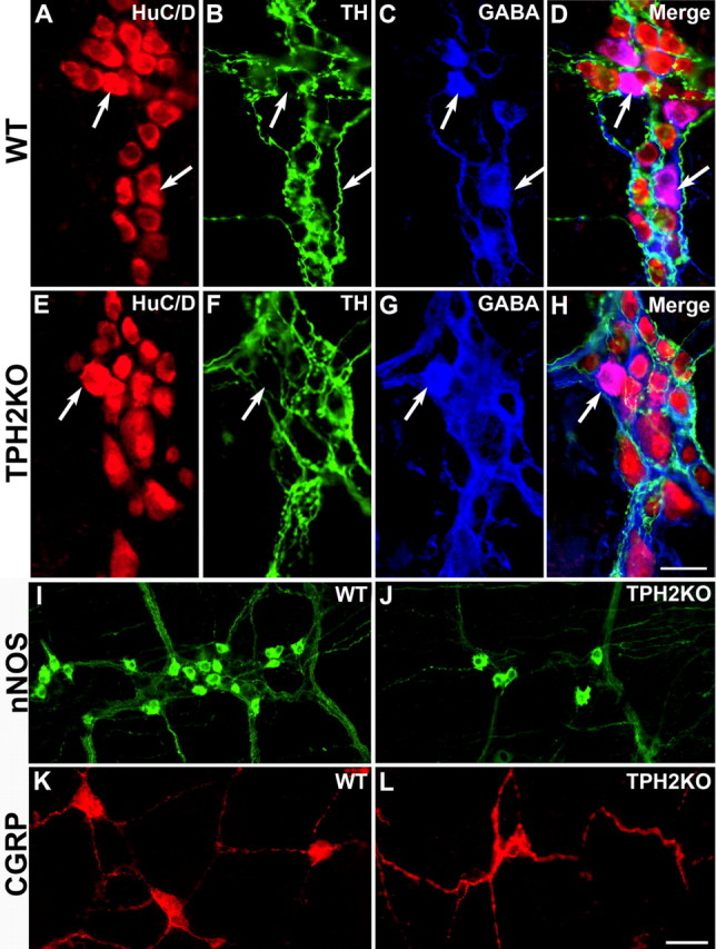
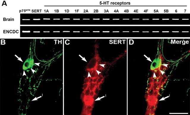
The gut contains a large 5-HT pool in enterochromaffin (EC) cells and a smaller 5-HT pool in the enteric nervous system (ENS). During development, enteric neurons are generated asynchronously. We tested hypotheses that serotonergic neurons, which arise early, affect development/survival of later-born dopaminergic, GABAergic, nitrergic, and calcitonin gene-related peptide-expressing neurons and are essential for gastrointestinal motility. 5-HT biosynthesis depends on tryptophan hydroxylase 1 (TPH1) in EC cells and on TPH2 in neurons; therefore, mice lacking TPH1 and/or TPH2 distinguish EC-derived from neuronal 5-HT. Deletion of TPH2, but not TPH1, decreased myenteric neuronal density and proportions of dopaminergic and GABAergic neurons but did not affect the extrinsic sympathetic innervation of the gut; intestinal transit slowed in mice lacking TPH2 mice, but gastric emptying accelerated. Isolated enteric crest-derived cells (ENCDCs) expressed the serotonin reuptake transporter (SERT) and 15 subtypes of 5-HT receptor. Addition of 5-HT to cultures of isolated ENCDCs promoted total and dopaminergic neuronal development. Rings of SERT-immunoreactive terminal axons surrounded myenteric dopaminergic neurons and SERT knock-out increased intestinal levels of dopamine metabolites, implying that enteric dopaminergic neurons receive a serotonergic innervation. Observations suggest that constitutive gastrointestinal motility depends more on neuronal than EC cell serotonin; moreover, serotonergic neurons promote development/survival of some classes of late-born enteric neurons, including dopaminergic neurons, which appear to innervate and activate in the adult ENS.


Figures

Figure 1.
Figure 1.
Total GI transit time, small intestine transit, and colonic motility are decreased but gastric emptying is accelerated in mice that lack TPH2 or TPH1 and TPH2. A, Carmine red was administered orally, and the time to development of red stools was noted. GI transit time in TPH1KO, TPH2KO, and TPH1/2dKO mice was compared, respectively, with that of wild-type littermates. Total GI transit time was increased (slower than littermates) in TPH2KO and TPH1/2dKO animals; however, the total GI transit time in TPH1KO mice did not differ from that of their wild-type littermates. Total GI transit time in TPH1/2dKO animals was not different from that in mice lacking only TPH2. B, Gastric emptying was measured after gavage of rhodamine dextran. The percentage of administered rhodamine dextran that emptied from the stomach in 15 min was determined in TPH1KO, TPH2KO, and TPH1/2dKO mice and compared with that of their respective wild-type littermates. The proportion of gastric contents that emptied was greater in TPH2KO and TPH1/2dKO animals than in the respective littermates; however, gastric emptying in TPH1KO mice did not differ from that in their wild-type littermates. Gastric emptying in TPH1/2dKO animals was not different from that in mice lacking only TPH2. C, Small intestinal transit was studied by measurement of geometric center of rhodamine dextran. The numbers of the geometric center, 1–10, represent slow to fast small intestine transit. Small intestinal transit was significantly slower in TPH2KO and TPH1/2dKO animals than in their respective wild-type littermates; however, small intestinal transit in TPH1KO mice did not differ significantly from that of their wild-type littermates. Small intestinal transit in TPH1/2dKO animals was not different from that in mice lacking only TPH2. D, Colonic motility was estimated by measuring the time required to expel a glass bead inserted into the rectum for a distance of 2 cm. This time was significantly greater (slower motility) in TPH2KO and TPH1/2dKO animals than in their respective wild-type littermates; however, the time to expel the bead in TPH1KO mice did not differ significantly from that in their wild-type littermates. The time required to expel the bead in TPH1/2dKO mice was not significantly different from that in mice lacking only TPH2. ns, Not significant.
Figure 2.
Figure 2.
Numbers of total and dopaminergic enteric neurons are decreased in mice that lack TPH2 or TPH1 and TPH2. A, The total number of neurons was obtained from counts made in whole mounts of dissected laminar preparations of longitudinal muscle with adherent myenteric plexus. The immunocytochemical detection of HuC/D was used as a neuronal marker. The numbers of total enteric neurons in TPH1KO, TPH2KO, and TPH1/2dKO mice were compared, respectively, with those of their wild-type littermates. Significantly fewer total neurons were found in TPH2KO and TPH1/2dKO animals than in their wild-type littermates; however, the total number of neurons in TPH1KO mice was not significantly different from that in their wild-type littermates. The number of neurons in TPH1/2dKO animals did not significantly differ from that in mice lacking only TPH2. B, TH was identified immunocytochemically and used as a marker for enteric dopaminergic neurons. Preparations were the same as used to investigate total neuronal numbers. Again, the numbers of TH-immunoreactive neurons in TPH1KO, TPH2KO, and TPH1/2dKO mice were compared with those of their respective wild-type littermates. The numbers of enteric dopaminergic neurons were significantly reduced in TPH2KO and TPH1/2dKO animals; however, the numbers of dopaminergic neurons in TPH1KO mice did not differ significantly from that of their wild-type littermates. The numbers of dopaminergic neurons in TPH1/2dKO animals was not significantly different from those in mice lacking only TPH2. Although both total and dopaminergic neurons were reduced in mice lacking TPH2 and TPH1 and TPH2, the deficit in dopaminergic neurons was more severe than that in total neurons. C, D, Neurons immunostained with antibodies to HuC/D in wild-type littermates (C) and TPH1/2dKO mice (D). E, F, Neurons immunostained with antibodies to TH in wild-type littermates (E) and TPH1/2dKO mice (F). G, H, Neurons immunostained with antibodies to HuC/D in wild-type littermates (G) and TPH2KO mice (H). I, J, Neurons immunostained with antibodies to TH in wild-type littermates (I) and TPH2KO mice (F). Neurons immunostained with antibodies to HuC/D in wild-type littermates (K) and TPH1KO mice (L). M, N, Neurons immunostained with antibodies to TH in wild-type littermates (M) and TPH1KO mice (N). Scale bars: L, 50 μm (for all HuC/D images); N, 25 μm (for all TH images). ns, Not significant.
Figure 3.
Figure 3.
Transcripts encoding TH and DAT are downregulated in the ileum of mice that lack TPH2 or TPH1 and TPH2. Real-time PCR was used to analyze TH and DAT expression quantitatively in the ileum. A, Expression of TH in TPH1KO, TPH2KO, and TPH1/2dKO mice was compared, respectively, with that in their wild-type littermates; data are expressed as percentage WT. Transcripts encoding TH were significantly less abundant in TPH2KO and TPH1/2dKO animals than in their wild-type littermates. The abundance of transcripts encoding TH in TPH1KO mice, however, did not differ significantly from that in their wild-type littermates. TH expression in TPH1/2dKO animals was not significantly different from that in mice lacking only TPH2. B, Expression of DAT in TPH1KO, TPH2KO, and TPH1/2dKO mice was compared, respectively, with that in their wild-type littermates; data are expressed as percentage WT. The abundance of transcripts encoding DAT in TPH2KO and TPH1/2dKO animals was significantly less than that in their wild-type littermates. The abundance of transcripts encoding DAT in TPH1KO mice, however, was greater than that in their wild-type littermates. DAT expression in TPH1/2dKO animals was not significantly different from that in mice lacking only TPH2. ns, Not significant.
Figure 4.
Figure 4.
5-HT stimulates the development and/or survival of total and dopaminergic enteric neurons in cultures of ENCDCs isolated from E16 fetal mouse gut. ENCDCs were immunoselected with antibodies to p75NTR and cultured for 6 d in the absence or presence of exogenous 5-HT (1 μm). The TPH inhibitor PCPA (1 μm) was added to the medium to prevent biosynthesis of endogenous 5-HT. A, TH was identified immunocytochemically and was used as a dopaminergic marker. The abundance of TH-immunoreactive neurons in 5-HT-treated cultures was significantly greater than that in control cultures exposed only to vehicle. B, HuC/D was identified immunocytochemically and was used as a neuronal marker. The abundance of neurons in 5-HT-treated cultures was significantly greater than in control cultures exposed only to vehicle. C, Although numbers of both total neurons and dopaminergic neurons increased after the addition of 5-HT, the proportion of neurons that were dopaminergic was not significantly different in 5-HT-treated cultures from that of control cultures exposed only to vehicle. D, E, HuC/D-immunoreactive neurons (red fluorescence) were found in small aggregates in both vehicle-treated (D) and 5-HT-treated (E) cultures. The numbers and the sizes of the aggregates, however, were both larger in 5-HT-treated cultures than in cultures exposed only to vehicle. F–I, Double-labeled immunocytochemistry was used to locate simultaneously the immunoreactivities of HuC/D and TH (green fluorescence). The cell bodies of TH-immunoreactive (dopaminergic) neurons were found within aggregates of HuC/D-immunoreactive neurons. F, Vehicle-treated culture. A single dopaminergic neuron appears within an aggregate of HuC/D-immunoreactive neurons. No dopaminergic neurites are visible. G, 5-HT-treated culture. Dopaminergic neurons are more numerous and extend TH-immunoreactive neurites that project to adjacent neuronal aggregates. H, I, 5-HT-treated cultures. H, Dopaminergic neurites project from neurons within an aggregate to the adjacent substrate, in which they terminate in a series of varicosities. I, Some dopaminergic neurites in 5-HT-treated cultures are very long and branch extensively. Scale bars: D, E, 100 μm; F–H, 20 μm; I, 50 μm. ns, Not significant.
Figure 5.
Figure 5.
Enteric levels of DA and HVA are higher in the ileum of SERTKO than in the ileum of WT littermates. HPLC with electrochemical detection was used to measure levels of DA, DOPAC, and HVA in the ileum, colon, and brain of SERTKO mice and their WT littermates. A, DA levels in neither the ileum nor the colon of SERTKO mice were significantly different from those in the ileum and colon of their wild-type littermates. B, The level of HVA was significantly greater than that of WT littermate controls in the ileum but not in the colon of SERTKO animals. C, The total level of DA plus its metabolites HVA and DOPAC was also significantly greater in the ileum of SERTKO mice than in their WT littermates, although again, the increase in the colon did not reach statistical significance. D, The level of 5-HT in neither the ileum nor colon of SERTKO mice differed significantly from that in the ileum and colon of wild-type littermate controls. E, The concentration of 5-HIAA, the 5-HT metabolite, in the colon was significantly lower in SERTKO mice than their WT littermates. F, In SERTKO mice, the total of 5-HT and 5-HIAA was significantly lower in both the ileum and colon than in WT animals. ns, Not significant.
Figure 6.
Figure 6.
Transcripts encoding SERT are downregulated in the ileum and colon of TPH1/2dKO mice. Real-time PCR was used to quantify to abundance of transcripts encoding SERT. For each sample, the abundance of transcripts encoding SERT was normalized to that of GAPDH. Data are expressed as the ratio of transcripts encoding SERT to those of GAPDH. SERT expression in both ileum and colon of TPH1/2dKO animals was significantly lower than that in their wild-type littermates.
Figure 7.
Figure 7.
Deletion of TPH2 reduces the numbers of GABAergic, nitrergic, and CGRP-expressing neurons but does not affect the density of sympathetic terminal axons in the ENS; the immunoreactivities of TH and GABA are not coincident. The immunoreactivities of HuC/D (total neurons), TH, and GABA were demonstrated simultaneously in LMMP preparations of WT (A–D) and TPH2KO (E–H) mice. A, WT mouse; HuC/D; B, WT mouse; TH. C, WT mouse; GABA. D, Merged image. E, TPH2KO mouse; HuC/D; F, TPH2KO mouse; TH. G, TPH2KO mouse; GABA. H, TPH2KO mouse; merged image. Whole-mount preparations were also used to demonstrate the immunoreactivities of nNOS in the myenteric (I, J) and CGRP in the submucosal plexus (K, L). I, WT mouse; nNOS. J, TPH2KO mouse; nNOS. K, WT mouse; CGRP. L, TPH2KO mouse; CGRP. Note that the density of TH-immunoreactive sympathetic terminals in the myenteric plexus is similar in WT (B) and TPH2KO (F) mice. Scale bars: A–H, 25 μm; I–L, 50 μm.
Figure 8.
Figure 8.
ENCDC precursor late-born enteric neurons express many subtypes of 5-HT receptor and SERT; myenteric dopaminergic neurons do not express SERT but are surrounded by a ring of SERT-immunoreactive (putatively serotonergic) terminal axons. A, Immunoselection with antibodies to p75NTR was used to isolate from the fetal intestine at E16. RT-PCR was used to determine whether these cells express transcripts encoding 15 subtypes, encompassing each of the families of 5-HT receptor. The brain was investigated as a positive control. In addition to p75NTR, both brain and ENCDCs contained transcripts encoding SERT, 5-HT1A, 5-HT1B, 15-HT1D, 5-HT1F, 5-HT2A, 5-HT2B, 5-HT3A, 5-HT4A, 5-HT4B, 5-HT4E, 5-HT4F, 5-HT5A, 5-HT5B, 5-HT6, and 5-HT7 receptors. B–D, TH and SERT immunoreactivities were demonstrated simultaneously in whole mounts of LMMP from adult mouse ileum. The location, in each panel of a TH-immunoreactive nerve cell body, is shown by an arrow, the location of the ring of SERT-immunoreactive terminal axons by arrowheads, and a SERT-immunoreactive nerve cell body by an arrow with an asterisk. B, TH immunoreactivity. C, SERT immunoreactivity. D, Merged image. Scale bar, 50 μm.

References

    1. Bertrand PP. Real-time detection of serotonin release from enterochromaffin cells of the guinea-pig ileum. Neurogastroenterol Motil. 2004;16:511–514. - PubMed
    1. Bertrand PP, Bertrand RL. Serotonin release and uptake in the gastrointestinal tract. Auton Neurosci. 2010;153:47–57. - PubMed
    1. Bertrand PP, Hu X, Mach J, Bertrand RL. Serotonin (5-HT) release and uptake measured by real-time electrochemical techniques in the rat ileum. Am J Physiol Gastrointest Liver Physiol. 2008;295:G1228–G1236. - PubMed
    1. Bian X, Patel B, Dai X, Galligan JJ, Swain G. High mucosal serotonin availability in neonatal guinea pig ileum is associated with low serotonin transporter expression. Gastroenterology. 2007;132:2438–2447. - PMC - PubMed
    1. Blaugrund E, Pham TD, Tennyson VM, Lo L, Sommer L, Anderson DJ, Gershon MD. Distinct subpopulations of enteric neuronal progenitors defined by time of development, sympathoadrenal lineage markers, and Mash-1-dependence. Development. 1996;122:309–320. - PubMed

Publication types

MeSH terms

LinkOut - more resources