Case Reports
. 2011 Oct;131(10):2017-25.
doi: 10.1038/jid.2011.157. Epub 2011 Jun 16.

Homozygosity mapping and whole-exome sequencing to detect SLC45A2 and G6PC3 mutations in a single patient with oculocutaneous albinism and neutropenia

Affiliations
Case Reports

Homozygosity mapping and whole-exome sequencing to detect SLC45A2 and G6PC3 mutations in a single patient with oculocutaneous albinism and neutropenia

Andrew R Cullinane et al. J Invest Dermatol. 2011 Oct.

Abstract

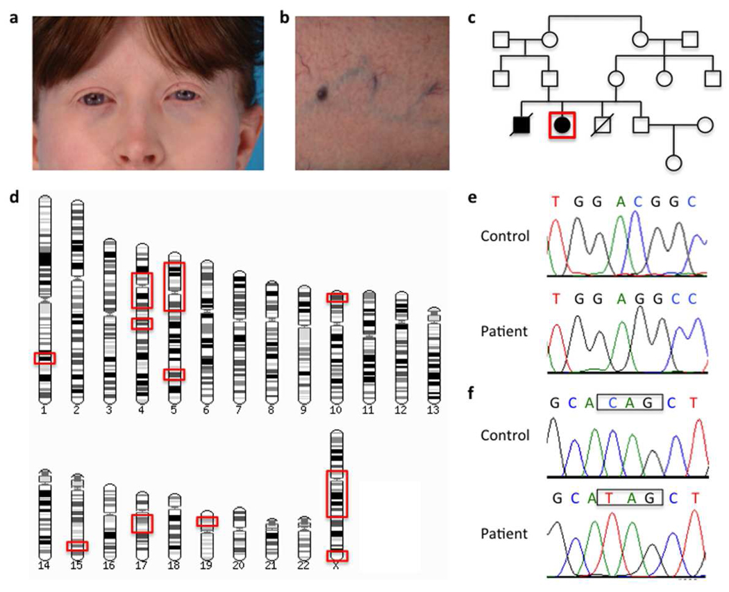
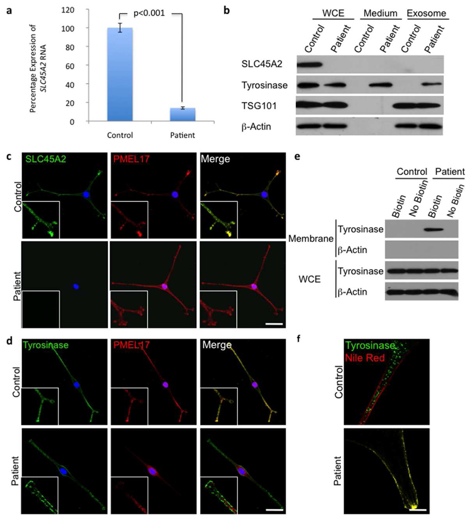
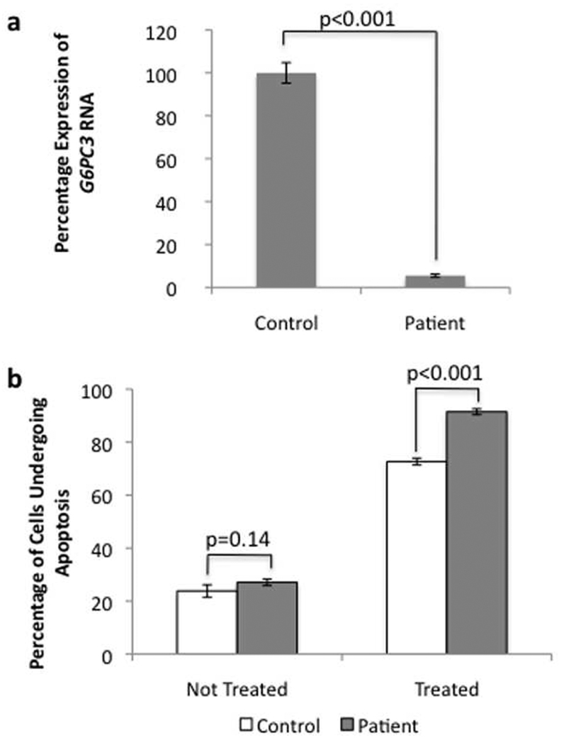
We evaluated a 32-year-old woman whose oculocutaneous albinism (OCA), bleeding diathesis, neutropenia, and history of recurrent infections prompted consideration of the diagnosis of Hermansky-Pudlak syndrome type 2. This was ruled out because of the presence of platelet δ-granules and absence of AP3B1 mutations. As parental consanguinity suggested an autosomal recessive mode of inheritance, we employed homozygosity mapping, followed by whole-exome sequencing, to identify two candidate disease-causing genes, SLC45A2 and G6PC3. Conventional dideoxy sequencing confirmed pathogenic mutations in SLC45A2, associated with OCA type 4 (OCA-4), and G6PC3, associated with neutropenia. The substantial reduction of SLC45A2 protein in the patient's melanocytes caused the mislocalization of tyrosinase from melanosomes to the plasma membrane and also led to the incorporation of tyrosinase into exosomes and secretion into the culture medium, explaining the hypopigmentation in OCA-4. Our patient's G6PC3 mRNA expression level was also reduced, leading to increased apoptosis of her fibroblasts under endoplasmic reticulum stress. To our knowledge, this report describes the first North American patient with OCA-4, the first culture of human OCA-4 melanocytes, and the use of homozygosity mapping, followed by whole-exome sequencing, to identify disease-causing mutations in multiple genes in a single affected individual.


Conflict of interest statement

Conflict of interest

The authors declare no conflict of interest.

Figures

Figure 1
Figure 1. Clinical findings and DNA analysis
a) Fair skin and classic red hair in our patient, typical for OCA-4 patients. b) Prominent superficial venous pattern seen in all previously described G6PC3 mutation-positive neutropenia patients. c) Pedigree showing affected and unaffected individuals; our patient is depicted by the red box. Note the consanguineous background of the family. d) Regions of homozygosity in the genome of the patient (shown within red boxes). e) Chromatogram of the mutation in SLC45A2 (c.986delC, p.T329RfsX68). f) Chromatogram of the mutation in G6PC3 (c.829C>T, p.Q277X). Control sequences are shown for comparison.
Figure 2
Figure 2. Functional analysis of the OCA-4 phenotype
a) Quantitative real-time PCR results for SLC45A2 mRNA expression in patient compared to control melanocytes. Values shown are percentage expression of SLC45A2 in patient cells compared to control cells, normalized by ACTB (Error bars = ± 1 SEM, n=3, p<0.001). b) Western blots of whole cell extracts (WCE), conditioned medium and exosome pellets from control and patient melanocytes. There is no detectable SLC45A2 protein expression and tyrosinase expression is reduced in patient cells compared with control cells. Tyrosinase is present in the medium from patient cells, suggesting that this protein is abnormally secreted from these cells; it is also found in the excreted exosome pellet. The media were collected from an equal number of cells and equal volumes of concentrated proteins were loaded onto the gel. Loading was controlled by assessing β-actin expression in the whole cell lysate of the corresponding cells. The western blot with the exosome marker TSG101 demonstrates that exosomes were isolated from the conditioned medium. c) Control and patient cells stained for SLC45A2 and the melanosome marker PMEL17, showing co-localization (inserts) of the 2 proteins in control cells. Consistent with the western blot, no staining for SLC45A2 is seen in patient cells but PMEL17 looks comparable to control cells. d) Control and patient cells stained for tyrosinase and PMEL17, show the co-localization (inserts) of the 2 proteins in control cells. No co-localization is seen in patient cells, and tyrosinase appears predominantly in the plasma membrane. Nuclei are stained with DAPI; scale bar represents 20 µm. e) Plasma membrane protein biotinylation assay. Only the patient’s cells showed tyrosinase on the plasma membrane. No β-Actin was detected in the membrane fraction (demonstrating purity). Whole cell lysates showed tyrosinase expression in control and patient’s cells (loading controlled by β-actin). f) High magnification images of control and patient cells stained for tyrosinase and the plasma membrane marker Nile Red. Tyrosinase membrane localization in the patient is evidenced by the co-localization of tyrosinase and Nile Red. Scale bar represents 5 µm.
Figure 3
Figure 3. Functional analysis of the G6PC3 phenotype
a) Quantitative real-time PCR results for G6PC3 mRNA expression in patients compared to control melanocytes. Values shown are percentage expression of G6PC3 in patient cells compared to control cells, normalized by ACTB (Error bars = ± 1 SEM, n=3, p<0.001). b) Flow cytometry results using the Annexin V apoptosis assay for control and patient cells with and without ER stress induction (Error bars = ± 1 SEM, n=3). Without 5mM dithiothreitol treatment no signicant difference can be seen; with treatment, there is a clear increase in apoptosis in patient cells.

Comment in

References

    1. Anikster Y, Huizing M, White J, et al. Mutation of a new gene causes a unique form of Hermansky-Pudlak syndrome in a genetic isolate of central Puerto Rico. Nat Genet. 2001;28:376–380. - PubMed
    1. Bentley DR, Balasubramanian S, Swerdlow HP, et al. Accurate whole human genome sequencing using reversible terminator chemistry. Nature. 2008;456:53–59. - PMC - PubMed
    1. Boissy RE, Zhao H, Oetting WS, et al. Mutation in and lack of expression of tyrosinase-related protein-1 (TRP-1) in melanocytes from an individual with brown oculocutaneous albinism: a new subtype of albinism classified as "OCA3". Am J Hum Genet. 1996;58:1145–1156. - PMC - PubMed
    1. Bonilla MA, Gillio AP, Ruggeiro M, et al. Effects of recombinant human granulocyte colony-stimulating factor on neutropenia in patients with congenital agranulocytosis. N Engl J Med. 1989;320:1574–1580. - PubMed
    1. Boztug K, Appaswamy G, Ashikov A, et al. A syndrome with congenital neutropenia and mutations in G6PC3. N Engl J Med. 2009;360:32–43. - PMC - PubMed

Publication types

MeSH terms