. 2011 Jun;13(6):504-15.
doi: 10.1593/neo.101590.

Insulin-like growth factor-dependent proliferation and survival of triple-negative breast cancer cells: implications for therapy

Affiliations

Insulin-like growth factor-dependent proliferation and survival of triple-negative breast cancer cells: implications for therapy

Zoë Davison et al. Neoplasia. 2011 Jun.

Abstract

Triple-negative breast cancers have a poor prognosis and are not amenable to endocrine- or HER2-targeted therapies. The prevailing view is that targeting the insulin-like growth factor (IGF) signal transduction pathway will not be beneficial for triple-negative breast cancers because their growth is not IGF-responsive. The present study investigates the importance of IGFs in the proliferation and survival of triple-negative breast cancer cells. Estrogen and progesterone receptors, HER2, type I IGF, and insulin receptors were measured by Western transfer analysis. The effects of IGF-1 on proliferation were assessed by DNA quantitation and on cell survival by poly (ADP-ribose) polymerase cleavage. The effect of IGF-1 on phosphorylation of the IGF receptors, Akt and mitogen-activated protein kinase, was measured by Western transfer analysis. Seven cell lines were identified as models of triple-negative breast cancer and shown to express IGF receptors at levels similar to those present in estrogen-responsive cell lines known to respond to IGFs. IGF-1 increased the proliferation and cell survival of all triple-negative cell lines. Proliferation was attenuated after reduction of type I IGF receptor expression. Cells that express higher levels of receptor were more sensitive to subnanomolar IGF-1 concentrations, but the magnitude of the effects was not correlated simply with the absolute amount or phosphorylation of the IGF receptors, Akt or mitogen-activated protein kinase. These results show that IGFs stimulate cell proliferation and promote cell survival in triple-negative breast cancer cells and warrant investigation of the IGF signal transduction pathway as a therapeutic target for the treatment of triple-negative breast cancer.


Figures

Figure 1
Figure 1
Expression of steroid and growth hormone receptors in cultured breast cells. BT-474, MCF-7, T-47D, ZR-75, EFF-3, EFM-19, BT-20, MDA-MB-231 (MDA-231), MDA-MB-468 (MDA-468), MDA-MB-435s (MDA-435s), Hs 578T, BT-549, HBL-100, SK-BR-3, MCF-10A, and HUMEC (HUM) cells were grown to 70% confluence in routine culture medium and lysed, and 20 µg of protein was electrophoresed on polyacrylamide gels and transferred to nitrocellulose as described in the Materials and Methods. Membranes were incubated with estrogen receptor (67 kDa), progesterone receptor (118 kDa), HER2 (185 kDa) (A), type I IGF receptor (95 kDa), or insulin receptor (95 kDa) (B) antibodies and developed by enhanced chemiluminescence as described in the Materials and Methods. Representative Western transfer images are shown on the left-hand side of the figure. The membranes were incubated secondly with a GAPDH antibody (data not shown), which confirmed equal loading and transfer. The analysis was repeated at least twice on four or more individual samples prepared from MCF-7, T-47D, ZR-75, EFF-3, EFM-19, MDA-MB-231, Hs 578T, BT-20, HBL-100, and SK-BR-3 breast epithelial cells and on three individual samples prepared from BT-474, MDA-MB-435s, MDA-MB-468, BT-549, MCF-10A, and HUMEC cells. The amount of each protein was determined by densitometric scanning of x-ray films followed by analysis with LabWorks 4.0 software and correcting for GAPDH as described in the Materials and Methods. The amount of each protein was expressed as the percentage of the amount of protein in the cell line that expresses themost of that protein and is represented in the histograms shown on the right hand side of the figure. Mean values ± SEM are shown. The filled columns indicate expression in triple-negative cell lines.
Figure 2
Figure 2
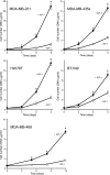
Stimulation of proliferation of triple-negative breast cancer cells by IGF-1. MDA-MB-231, MDA-MB-435s, Hs 578T, and BT-549 cells were plated in 16-mm-diameter tissue culture wells, allowed to attach, withdrawn from the growth stimulatory effects of normal culture medium, and then cultured for the indicted lengths of time in the absence (○) or presence (●) of 50 ng/ml IGF-1 as described in the Materials and Methods. MDA-MB-468 cells were plated in Corning CellBIND plates. Cells were lysed, and the DNA content of the wells was measured. Bars, SEM. Asterisks show times at which there were significantly more cells in the presence of IGF-1 than in its absence.
Figure 3
Figure 3
Effect of decreased expression of type I IGF receptor on cell proliferation in MDA-MB-231 and Hs 578T cells. MDA-MB-231 (A) and Hs 578T (B) cells were transfected with shRNA plasmid pKD-IGF-IR-v2 and then cultured for 3 days in the absence or presence of 50 ng/ml IGF-1. Cells were lysed, and 20 µg of protein was electrophoresed on polyacrylamide gels and transferred to nitrocellulose. Membranes were incubated with type I IGF receptor (95 kDa) antibody and developed by enhanced chemiluminescence as described in the Materials and Methods. Representative Western transfer images after exposure to the x-ray film for 30 seconds (A) and 3 hours (B) are shown. The membranes were incubated secondly with a GAPDH antibody (data not shown), which confirmed equal loading and transfer. MDA-MB-231 (C) and Hs 578T (D) cells were transfected with shRNA plasmid pKD-IGF-IR-v2 and then cultured in the absence or presence of 50 ng/ml IGF-1 as described in the Materials and Methods. Cells were lysed, and the DNA content of the wells was measured. Bars, SEM. Asterisks show times at which there were significantly fewer cells grown in the presence of 50 ng/ml IGF-1 in the wells that contained cells transfected with pKD-IGF-IR-v2 than in wells that contained cells transfected with the control plasmid (t test, P < 0.05).
Figure 4
Figure 4
Protective effect of IGF-1 on programmed cell death in triple-negative breast cancer cells. MDA-MB-231, HBL-100, BT-549, MDA-MB-468, Hs 578T, BT-20, and MDA-MB-435s cells were cultured and treated with staurosporine in the absence and presence of 50 ng/ml IGF-1 as described in the Materials and Methods. Cells were lysed after various times, and the amount of PARP cleavage was measured by Western transfer analysis. Representative Western transfer images are shown in A. The positions of cleaved and uncleaved PARP proteins are indicated on the right of the images. The amount of each protein was determined by densitometric scanning of the x-ray films followed by analysis with LabWorks 4.0 software. In B, the amount of cleaved 89-kDa form of PARP protein expressed as a percentage of total PARP protein is shown in the absence (Ë) or presence (Ï) of IGF-1. Bars, SEM. Asterisks show PARP cleavage that is statistically significantly less in the presence of IGF-1 than in its absence.
Figure 5
Figure 5
Phosphorylation of IGF receptors, Akt and MAPK, in response to IGF-1 in triple-negative breast cancer cells. BT-20, MDA-MB-231, MDA-MB-468, MDA-MB-435s, Hs 578T, BT-549, HBL-100, and SK-BR-3 cells were seeded in routine culture medium, cultured for 2 days in phenol red-free medium supplemented with charcoal-treated serum, for 2 hours in serum-free medium, and then stimulated with 50 ng/ml IGF-1 for 15 minutes in the same medium, and protein lysates were prepared. Aliquots of 10 µg of protein were electrophoresed on a polyacrylamide gel; transferred to nitrocellulose; incubated with phospho-IGF receptor, phospho-Akt, or phospho-ERK1/2 antibodies; and developed using enhanced chemiluminescence as described in the Materials and Methods. Because we detected exceptionally high amounts of phosphorylated Akt and MAPK in BT-20 cells and of phosphorylated MAPK in HBL-100 cells, lower exposures of the membranes to the x-ray films are shown for these proteins. The membranes were incubated secondly with a tubulin antibody to confirm equal loading and transfer. The protein samples were analyzed to measure the total level of the proteins and to confirm that the levels do not change during the experiment. Representative images of the results obtained are shown underneath the panels of the corresponding phosphorylated proteins.
Figure 6
Figure 6
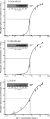
Stimulation of phosphorylation of IGF receptors by different concentrations of IGF-1. MDA-MB-231 (A), MDA-MB-468 (B), and Hs 578T (C) cells were withdrawn for 2 days and then incubated in the presence of the indicated concentrations of IGF-1 for 15 minutes. Cells were lysed, and phosphorylation of IGF receptors was measured and quantified as described in the Materials and Methods. Representative images from the membranes after exposure to an x-ray film for 1 minute (MDA-MB-231), 10 minutes (MDA-MB-468), and 5 hours (Hs 578T) are shown as inserts. The amount of phosphorylated receptor was determined by densitometric scanning of the x-ray films followed by analysis with LabWorks 4.0 software. Bars, SEM.
Figure 7
Figure 7
Stimulation of cell proliferation and cell survival by different concentrations of IGF-1. MDA-MB-231 (A) and Hs 578T (B) cells were withdrawn from growth stimuli and cultured in the presence of the indicated concentrations of IGF-1. Cells were lysed, and the DNA content of the wells was measured as described in the Materials and Methods. MDA-MB-231 (C) and Hs 578T (D) cells were treated with staurosporine in the absence or presence of the indicated concentrations of IGF-1. Cells were lysed, and the percentage of PARP cleavage was determined as described in the Materials and Methods. Bars, SEM. Asterisks indicate the lowest concentration of IGF-1 at which a significant effect of IGF-1 was detected.

References

    1. Schneider BP, Winer EP, Foulkes WD, Garber J, Perou CM, Richardson A, Sledge GW, Carey LA. Triple-negative breast cancer: risk factors to potential targets. Clin Cancer Res. 2008;14(24):8010–8018. - PubMed
    1. Reis-Filho JS, Tutt AN. Triple negative tumours: a critical review. Histopathology. 2008;52(1):108–118. - PubMed
    1. Rakha EA, Reis-Filho JS, Ellis IO. Basal-like breast cancer: a critical review. J Clin Oncol. 2008;26:2568–2581. - PubMed
    1. Bauer KR, Brown M, Cress RD, Parise CA, Caggiano V. Descriptive analysis of estrogen receptor (ER)-negative, progesterone receptor (PR)-negative, and HER2-negative invasive breast cancer, the so-called triple-negative phenotype: a population-based study from the California Cancer Registry. Cancer. 2007;109(9):1721–1728. - PubMed
    1. Stead LA, Lash TL, Sobieraj JE, Chi DD, Westrup JL, Charlot M, Blanchard RA, Lee JC, King TC, Rosenberg CL. Triple negative breast cancers are increased in black women regardless of age or body mass index. Breast Cancer Res. 2009;11(2):R18. - PMC - PubMed

Publication types

MeSH terms

LinkOut - more resources