

Intraoperative changes in idiopathic macular holes by spectral-domain optical coherence tomography
- PMID: 21677882
- PMCID: PMC3104860
- DOI: 10.1159/000328752
Intraoperative changes in idiopathic macular holes by spectral-domain optical coherence tomography
Abstract
Purpose: To examine anatomical changes in idiopathic macular holes during surgery using handheld spectral-domain optical coherence tomography (SD-OCT).
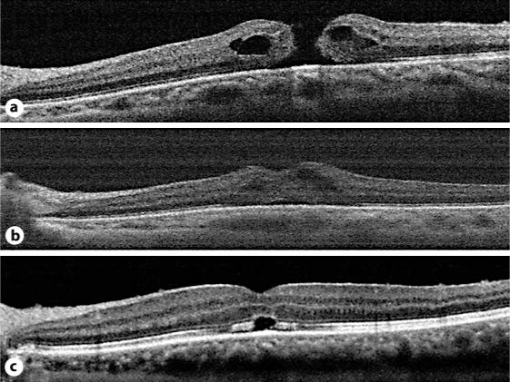
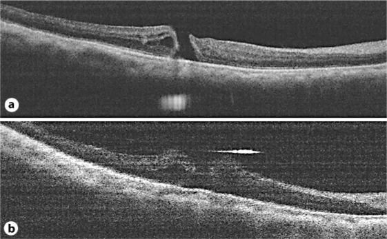
Methods: Five eyes of 5 patients who underwent surgery for the repair of idiopathic macular holes were examined. The surgery included standard 25-gauge, 3-port pars plana vitrectomy, removal of the internal limiting membrane (ILM), fluid-air exchange, and 20% sulfur hexafluoride tamponade. Intraoperative SD-OCT images of the macular holes were obtained after ILM removal and under fluid-air exchange using a handheld SD-OCT. From SD-OCT images, the macular hole base diameter (MHBD) was measured and compared.
Results: All macular holes were successfully closed after the primary surgery. The mean MHBD under fluid-air exchange was significantly smaller than the mean MHBD after ILM removal and the preoperative mean MHBD. In 1 eye with a stage 3 macular hole, SD-OCT images revealed that the inner edges of the macular hole touched each other under fluid-air exchange.
Conclusion: Fluid-air exchange significantly reduced MHBD during surgery to repair macular holes. Fluid-air exchange may be an important step for macular hole closure as it reduces the base diameter of the macular hole.
Keywords: Base diameter; Intraoperative OCT; Macular hole; Spectral-domain OCT.
Figures
References
-
- Binder S, Falkner-Radler CI, Hauger C, Matz H, Glittenberg C. Feasibility of intrasurgical spectral-domain optical coherence tomography. Retina. 2011 Epub ahead of print. - PubMed
-
- Wykoff CC, Berrocal AM, Schefler AC, Uhlhorn SR, Ruggeri M, Hess D. Intraoperative OCT of a full-thickness macular hole before and after internal limiting membrane peeling. Ophthalmic Surg Lasers Imaging. 2010;41:7–11. - PubMed
-
- Masuyama K, Yamakiri K, Arimura N, Sonoda Y, Doi N, Sakamoto T. Posturing time after macular hole surgery modified by optical coherence tomography images: a pilot study. Am J Ophthalmol. 2009;147:481–488. - PubMed
Publication types
LinkOut - more resources
Full Text Sources
Miscellaneous
