. 2011 Oct;112(10):2992-3001.
doi: 10.1002/jcb.23222.

Identification of a novel effector domain of BIN1 for cancer suppression

Affiliations

Identification of a novel effector domain of BIN1 for cancer suppression

Greta L Lundgaard et al. J Cell Biochem. 2011 Oct.

Abstract

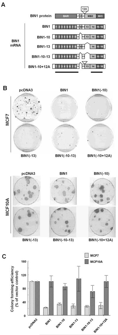
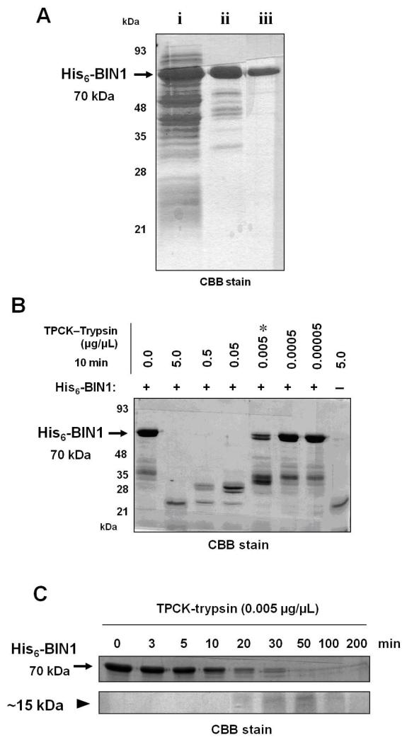
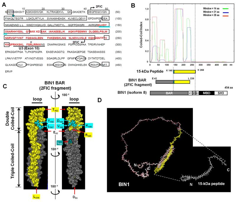
Bridging integrator 1 (BIN1) is a nucleocytoplasmic adaptor protein with tumor suppressor properties. The protein interacts with and inhibits the c-MYC transcription factor through the BIN1 MYC-binding domain (MBD). However, in vitro colony formation assays have clearly demonstrated that the MBD is not essential for BIN1-mediated growth arrest. We hypothesized that BIN1 contains a MYC-independent effector domain (MID) for cancer suppression. Because a functionally unique domain frequently contains a distinct structure, the human full-length BIN1 protein was subjected to limited trypsin digestion and the digested peptides were analyzed with Edman sequencing and mass spectrometry. We identified a trypsin-resistant peptide that corresponds to amino acids 146-268 of BIN1. It encompassed part of the BAR region, a putative effector region of BIN1. Computational analysis predicted that the peptide is very likely to exhibit coiled-coil motifs, implying a potential role for this region in sustaining the BIN1 structure and function. Like MBD-deleted BIN1, the trypsin-resistant peptide of BIN1 was predominantly present in the cytoplasm and was sufficient to inhibit cancer growth, regardless of dysregulated c-MYC activity. Our results suggest that the coiled-coil BIN1 BAR peptide encodes a novel BIN1 MID domain, through which BIN1 acts as a MYC-independent cancer suppressor.


Figures

Figure 1
Figure 1. Effects of BIN1 spliced isoforms on the colony forming activities of MCF7 and MCF10A cell lines
(A) Alternatively spliced isoforms of BIN1. The BIN1 protein is encoded by 16 exons. The BAR region is encoded by exons 1–8, U1 is encoded by exon 9, U3 is encoded by exon 10, U2 is encoded by exon 11, MBD is encoded by exons 13–14, and the SH3 domain is encoded by exons 15–16. Exon 12A has been detected in brain-specific transcripts (Wechsler-Reya et al., 1997) and metastatic melanoma cells (Ge at al. 1999). Horizontal bars indicate the common regions among the spliced isoforms. (B) Morphologies of MCF7 breast cancer and MCF10A untransformed breast epithelial cell lines after the formation of G418-resistant colonies. Growing MCF7 cells and MCF10A cells were stably transfected with the G418-resistant gene-expression vectors that encode indicated cDNAs of the human BIN1 splice isoforms, selected for 14 days and 21 days in G418 (500 μg/ml and 600 μg/ml)-containing growth medium, respectively, and stained with 4% (v/v) Giemsa (Invitrogen). (C) Giemsa-stained colonies of indicated cell lines were scored. The numbers of colonies transfected with the empty control vector were used as the positive control. The bars represent the means of three independent experiments ± SD.
Figure 2
Figure 2. Optimization of limited tryptic digestion of the human recombinant BIN1 protein, His6-BIN1
(A) A 30 μl aliquot of each fraction was subjected to 10% SDS-PAGE and stained with CBB R-250. The arrow indicates His6-BIN1 (~70 kDa). i) Escherichia coli lysates containing approximately 70 μg of crude protein; ii) post-Ni column fraction, containing approximately 15 μg of protein; and iii) post-Sephacryl S-200 gel filtration fraction, containing approximately 5 μg of protein. (B) Range-finding experiments for tryptic digestion of His6-BIN1 protein confirmed that 0.005 μg/μl (asterisk) was the optimal concentration of TPCK-trypsin for limited trypsinization of His6-BIN1 protein. (C) Time-course experiments for the tryptic digestion (0.005 μg/μl) of His6-BIN1 protein. The arrow and arrowhead indicate His6-BIN1 (approx. 70 kDa) and a BIN1 small peptide resistant to trypsin (approximately 15 kDa), respectively. Each aliquot was subjected to 14% SDS-PAGE and CBB staining. The procedures to purify the 15-kDa band were repeated at least three times to confirm the chromatographic reproducibility.
Figure 3
Figure 3. Identification and characterization of a BIN1 15-kDa peptide registrant to limited trypsin digestion
(A) Secondary structure prediction of BIN1. The PSIPRED program was used for the secondary structure prediction (McGuffin et al., 2000). The predicted helices and β-strands are presented as rectangles and ellipses, respectively. The region encoding the 15-kDa peptide is highlighted in red. The region indicated by arrows (E42–L239) corresponds to part of the human BIN1 BAR fragment, the three-dimensional structure of which is available online under the Protein Data Bank (PDB) accession number 2FIC (Casal et al., 2006). Leucine55 (L55), highlighted in a dark green circle, is a microsatellite mutation detected in a set of prostate cancer tissues (Ge et al., 2000). The KLVDY sequence necessary for BIN1-mediated anti-transforming activity is underlined in turquoise (DuHadaway et al., 2001). The region encoding U3 (or exon 10) is underlined in black. Arginine263 (asterisk) is the calculated C-terminal residue of the 15-kDa peptide fragment. (B) Prediction of the coiled-coil region in BIN1. The COILS server (Lupas et al., 1991) was used to predict the coiled-coil regions. The x-axis depicts the amino acid positions in the human BIN1 (isoform 8) protein and the y-axis shows the possibility of coiled-coil regions. The color denotes the window size used in the search. Three BIN1 polypeptides, the 15-kDa peptide (yellow), the BIN1 BAR (2FIC) fragment (Casal et al., 2006), and full-length BIN1 (isoform 8; 454 amino acids) (Sakamuro et al., 1996) were aligned. The KLVDY sequence is highlighted in turquoise. (C) The three-dimensional structure of the 15-kDa peptide (yellow) was modeled based on the structure of the human BIN1 BAR fragment (PDB accession number 2FIC), starting from E42 through to L239 (Casal et al., 2006). The KLVDY sequence, which is the N-terminus of the 15-kDa peptide fragment, is highlighted in turquoise. Double and triple coiled-coil domains are indicated with vertical bars, and loops with horizontal bars. The L55 residue in dark green corresponds to the location of a satellite mutation found in a set of patients with prostate cancer, as described previously (Ge et al., 2000). Residues D93, T162, and L239 are expected to exist in the hinge regions. (D) LOMETS (Local Meta-Threading-Server) program (Wu and Zhang, 2007) was used to predict the 3-D structure of the full-length BIN1 protein (left) and the 15-kDa peptide (right).
Figure 4
Figure 4. The 15-kDa peptide encodes the BIN1 MYC-independent effector domain (MID) for cancer suppression
(A) Colony formation suppression assays. LNCaP and MCF cancer cell lines were transfected with CMV-BIN1 (2.0 μg) or CMV-HA-Trp (2.0 μg). At 24 h after transfection, the cells were trypsinized and reseeded at a low density in standard growth medium containing G418 at 500 μg/ml. G418-resistant colonies of each cell line were stained and scored after two weeks of drug selection. The data presented are the percentages of colonies formed in the presence of G418 compared to the empty vectors. The bars represent the means of three experiments ± SD. (B) The EMS-Luc vector is a c-MYC-responsive reporter construct that contains four tandem repeats of the E-box sequence (small open dotes) followed by the SV40 minimal promoter (top). EMS–Luc (1.0 μg) reporter vector was cotransfected with 1.0 μg of pLPC empty vector, pLPC-BIN1, pLPC-BIN1ΔMBD, or pLPC-HA-Trp in proliferating LNCaP cells. The cells were harvested 48 h after transfection for the luciferase assays (bottom). Three independent transfections were performed for statistical analysis. (C) In situ immunofluorescence microscopy verified the cytoplasmic localization of transfected HA-BIN1ΔMBD and HA-Trp polypeptides and the nuclear localization of transfected BIN1 and endogenous PARP1 proteins in two BIN1-deficient cancer cell lines, MCF7 and DU145.

References

    1. Baker SJ, Markowitz S, Fearon ER, Willson JK, Vogelstein B. Suppression of human colorectal carcinoma cell growth by wild-type p53. Science. 1990;249(4971):912–915. - PubMed
    1. Brzovic PS, Meza J, King MC, Klevit RE. The cancer-predisposing mutation C61G disrupts homodimer formation in the NH2-terminal BRCA1 RING finger domain. J Biol Chem. 1998;273(14):7795–7799. - PubMed
    1. Casal E, Federici L, Zhang W, Fernandez-Recio J, Priego EM, Miguel RN, DuHadaway JB, Prendergast GC, Luisi BF, Laue ED. The crystal structure of the BAR domain from human BIN1/amphiphysin II and its implications for molecular recognition. Biochemistry. 2006;45(43):12917–12928. - PMC - PubMed
    1. Cassimere EK, Pyndiah S, Sakamuro D. The C-MYC-interacting proapoptotic tumor suppressor BIN1 is a transcriptional target for E2F1 in response to DNA damage. Cell Death & Diff. 2009;16(12):1641–1653. - PubMed
    1. DuHadaway JB, Sakamuro D, Ewert DL, Prendergast GC. BIN1 mediates apoptosis by C-MYC in transformed primary cells. Cancer Res. 2001;61(7):3151–3156. - PubMed

Publication types

MeSH terms

LinkOut - more resources