. 2011 Aug;29(8):1206-18.
doi: 10.1002/stem.674.

Optic vesicle-like structures derived from human pluripotent stem cells facilitate a customized approach to retinal disease treatment

Affiliations

Optic vesicle-like structures derived from human pluripotent stem cells facilitate a customized approach to retinal disease treatment

Jason S Meyer et al. Stem Cells. 2011 Aug.

Abstract

Differentiation methods for human induced pluripotent stem cells (hiPSCs) typically yield progeny from multiple tissue lineages, limiting their use for drug testing and autologous cell transplantation. In particular, early retina and forebrain derivatives often intermingle in pluripotent stem cell cultures, owing to their shared ancestry and tightly coupled development. Here, we demonstrate that three-dimensional populations of retinal progenitor cells (RPCs) can be isolated from early forebrain populations in both human embryonic stem cell and hiPSC cultures, providing a valuable tool for developmental, functional, and translational studies. Using our established protocol, we identified a transient population of optic vesicle (OV)-like structures that arose during a time period appropriate for normal human retinogenesis. These structures were independently cultured and analyzed to confirm their multipotent RPC status and capacity to produce physiologically responsive retinal cell types, including photoreceptors and retinal pigment epithelium (RPE). We then applied this method to hiPSCs derived from a patient with gyrate atrophy, a retinal degenerative disease affecting the RPE. RPE generated from these hiPSCs exhibited a disease-specific functional defect that could be corrected either by pharmacological means or following targeted gene repair. The production of OV-like populations from human pluripotent stem cells should facilitate the study of human retinal development and disease and advance the use of hiPSCs in personalized medicine.


Conflict of interest statement

COMPETING INTERESTS STATEMENT

The authors declare competing financial interests: J.A.T. is a founder, stockowner, consultant and board member of Cellular Dynamics International (CDI). He also serves as scientific advisor to and has financial interests in Tactics II Stem Cell Ventures. CDI currently has no products related to the retina. The other authors have no financial interests to disclose.

Figures

Fig. 1
Fig. 1. Isolation of optic vesicle-like structures from hESCs
Between day 20–25 of hESC differentiation, free-floating cell aggregates displayed two morphologies: phase-bright vesicle-like structures (arrows) or darker, rosetted spheres (arrowheads) (A). These populations were manually separated into vesicle-like (B) and nonvesicular (C) pools and independently cultured (arrows indicate rosettes in nonvesicular spheres). The definitive retinal progenitor marker CHX10 was expressed exclusively in the population of vesicle-like structures (DF), whereas ISLET-1 was initially expressed solely in the nonvesicular sphere population (GI). PCR analysis identified optic vesicle (left) and early forebrain (middle) transcription factors that were differentially expressed at day 20–25 in vesicle-like structures (Vesc) and nonvesicular spheres (Non Vesc), as well as factors that were present in both populations (right) (J). Immunostained sections of day 20 vesicle-like structures revealed co-expression of CHX10 and Ki67 (K). By day 50, many vesicle-like structures lost their distinctive morphology and developed internal rosettes (L), but remained CHX10+/Ki67+ (M).
Fig. 2
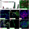
Fig. 2. Optic vesicle-like structures produce major neuroretinal cell types in a developmentally appropriate sequence
qPCR analysis of isolated OV-like structureswas performed every 10 days from day 20–120 to follow expression of genes indicative of selected neuroretinal cell types (data shown are relative to day 20 for each gene) (A). ICC was used to corroborate neuroretinal cell type identity at day 80: retinal ganglion cells (B), amacrine/horizontal cells (C), bipolar cells (D), and photoreceptors (EH).
Fig. 3
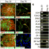
Fig. 3. Early forebrain spheres produce multiple neural cell types
After 20 days of differentiation, EFB spheres expressed βIII-TUBULIN (AC), PAX6 (A), SOX1 (B), and OTX2 (C). After 70 days, nearly all cells expressed markers of mature neurons or glia, including GABA, βIII-TUBULIN (D), tyrosine hydroxylase (TH), MAP2 (E), and GFAP (F). PCR analysis (G) at days 20 and 70 confirmed changes in transcription factor expression over time.
Fig. 4
Fig. 4. Photoreceptor-like cells from optic vesicle-like structures display a characteristic electrophysiological signature
Cells undergoing electrophysiological analysis were loaded with sulphorhodamine (A) and later immunostained to confirm photoreceptor marker expression (B). Expression of multiple genes involved in phototransduction was determined at day 80 of differentiation (C). (D) Average current density measured from 15 photoreceptor-like cells (solid circles) and 3 non-photoreceptor cells (open squares) plotted against applied step voltage. (E) Ionic current responses measured at +40 and +20 mV from a representative voltage-clamped cell before (control) and after (+TEA) application of TEA. TEA sensitive outward currents were calculated by mathematical subtraction. (F) I-V curve showing average steady state current amplitudes of the photoreceptor-like cells before (solid circles) and after (solid squares) bath application of TEA. (G) Time course of the effect of 8-br-cGMP on current amplitude in a representative photoreceptor-like cell. (H) Ionic current traces obtained in response to voltage pulses delivered before (control) and during (+8-br-cGMP) administration of 8-br-cGMP. The horizontal line represents zero current. (I) I-V curve showing average current responses before (solid circles) and during (solid squares) 8-br-cGMP administration. Results shown are an average of ≥4 experiments ± SEM.
Fig. 5
Fig. 5. Optic vesicle-like structures can be directed to an RPE fate
RPE was rarely observed in isolated OV-like structures after 50 days of differentiation (A). With the addition of Activin A between day 20–40, a subset of these structures became pigmented (B), whereupon they could be manually isolated and cultured separately (C). Plated pigmented structures were grown in the presence of FGF2, EGF, and heparin to promote outgrowth of cells (D). Upon removal of mitogens, RPE adopted its typical appearance (E) and expressed characteristic markers (F). Activin A-treated OV-like structures expressed higher levels of RPE-associated genes (G) and lower levels of neuroretinal-associated genes (H) by qPCR. Monolayers of RPE (I) were loaded with Fura-2 AM and stimulated with ATP while being monitored via epiflourescence imaging (J) to record changes in [Ca2+]i over time (K). Panel J is an epiflourescence image of the cells shown in panel I. Cells identified in panel J were used to generate the tracing in panel K. The bar in panel K shows the timing of ATP administration.
Fig. 6
Fig. 6. Human iPSC lines produce optic vesicle-like structures indistinguishable from hESCs
Variability was observed by qPCR in the expression of anterior neural/eye field genes at day 10 of differentiation across multiple hESC and hiPSC lines (A–B and Supporting Information Fig. S4), with low-expressing lines adopting a non-neural, epithelial-like identity (C–D). Higher relative expression levels of DKK1 (E) and, to a lesser extent, NOGGIN (F), at day 2 correlated with higher relative expression levels of anterior neural/eye field genes at day 10. OV-like structures generated from hiPSCs (G) uniformly expressed CHX10 (H) after 20–25 days. Acquisition of an anterior neural/eyefield fate at day 10 was enhanced through the addition of DKK1 and NOGGIN from day 2–4, as determined by qPCR (data expressed relative to untreated cultures) (I). DKK1-and NOGGIN-treated hiPSC cultures yielded colonies at day 10 with neuroepithelial morphology (J) that expressed anterior neural/eye field markers (K). FACS analysis demonstrated increased expression of PAX6 (L) and CHX10 (M) after 10 and 20 days of differentiation, respectively, in DKK1- and NOGGIN-treated hiPSC cultures.
Fig. 7
Fig. 7. Differentiation and testing of hiPSCs derived from a patient with gyrate atrophy
Fibroblasts (A) from a patient with gyrate atrophy (GA) due to an A226V mutation in OAT were reprogrammed to hiPSCs (B). (A226)OAT hiPSCs formed neural rosettes (C) at day 10 of differentiation that expressed anterior neural/eye field markers (D). Further maturation yielded multiple retinal cell types, including photoreceptor-like cells (E) and RPE (F). OAT activity in RPE derived from (A226)OAT hiPSCs was significantly less than that of control RPE from human prenatal eyes, hESCs, and hiPSCs (G). OAT enzyme activity in (A226)OAT hiPSC-RPE was restored in the presence of 600 μM vitamin B6 (H), or following gene repair via BAC-mediated homologous recombination (I). **p<0.0005; ***p<0.0001. Panels GI are representative examples of 3 independent experiments.

References

    1. Keller G. Embryonic stem cell differentiation: emergence of a new era in biology and medicine. Genes Dev. 2005;19:1129–1155. - PubMed
    1. Pera MF, Trounson AO. Human embryonic stem cells: prospects for development. Development. 2004;131:5515–5525. - PubMed
    1. Ebert AD, Yu J, Rose FF, Jr, et al. Induced pluripotent stem cells from a spinal muscular atrophy patient. Nature. 2009;457:277–280. - PMC - PubMed
    1. Gamm DM, Meyer JS. Directed differentiation of human induced pluripotent stem cells: a retina perspective. Regen Med. 2010;5:315–317. - PubMed
    1. Jin ZB, Okamoto S, Osakada F, et al. Modeling retinal degeneration using patient-specific induced pluripotent stem cells. PLoS One. 2011;6:e17084. - PMC - PubMed

Publication types

MeSH terms