. 2011 Jun 16;13(3):R90.
doi: 10.1186/ar3365.

Hyaluronan modulates accumulation of hypoxia-inducible factor-1 alpha, inducible nitric oxide synthase, and matrix metalloproteinase-3 in the synovium of rat adjuvant-induced arthritis model

Affiliations

Hyaluronan modulates accumulation of hypoxia-inducible factor-1 alpha, inducible nitric oxide synthase, and matrix metalloproteinase-3 in the synovium of rat adjuvant-induced arthritis model

Li-Wei Chou et al. Arthritis Res Ther. .

Abstract

Introduction: Hypoxia is a feature of the inflamed synovium in rheumatoid arthritis (RA). Intra-articular injection of hyaluronan (HA) may be considered a potential way to treat RA. However, the exact molecular mechanism of HA on decreased cellular responses to hypoxic environment is unclear. The present study has been designed to use the adjuvant-induced arthritis model to examine the effects of HA on the changes of immunohistochemical expressions of hypoxia-inducible factor-1alpha (HIF-1alpha), inducible nitric oxide synthase (iNOS), and matrix metalloproteinase-3 (MMP3) in the synovial tissues at the early phase of arthritic inflammation.

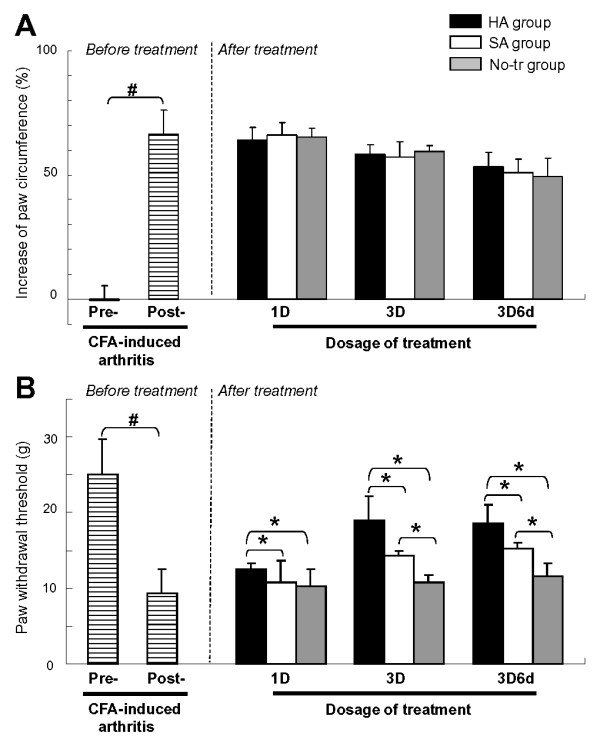
Methods: Monoarthritis was induced in adult male Sprague-Dawley (250-300 g) via intraarticular injection of complete Freund's adjuvant (CFA) into the tibiotarsal joint. The CFA-induction arthritis animals were divided into three groups: treatment (intraarticular injection of HA), placebo (intraarticular injection of saline) and controls (no treatments). Functional evaluations of edema and pain behavior, histology, and HIF-1alpha, iNOS, and MMP3 immunohistochemistry were performed before, after the first injection, three injections, and on the follow-up injection of the treatments.

Results: Intra-articular injection of HA also significantly suppressed the mechanical allodynia (p < 0.001) and overexpressions of HIF-1alpha (p < 0.001), iNOS (p = 0.004) and MMP3 (p < 0.001) immunoreactivity in synovium.

Conclusions: This study demonstrated that early intervention of HA is an effective protection against accumulation of inflammation-induced HIF-1alpha, iNOS, and MMP3 to limit erosive damage in CFA-induced model of arthritis.


Figures

Figure 1
Figure 1
Experimental design of the sequence of events for the entire course of the experiment. After evaluations that included measurements of paw edematous swelling and pain threshold, the animals were sacrificed for histology and immunohistochemistry. 1D, one dose; 3D, three doses; 3D6d, follow-up at the 6th day after three doses; CFA, complete Freund's adjuvant; HA, hyaluronan; No-tr, no treatment; SA, saline.
Figure 2
Figure 2
Results of edema (a) and pain behavioral (b) assessments. Data were calculated before treatment at the conditions of pre-complete Freund's adjuvant (CFA)-induced and post-CFA-induced arthritis and after treatment at conditions of one dose (1D), three doses (3D), and follow-up at the 6th day after three doses (3D6d) in hyaluronan (HA), saline (SA), and 'no treatment' (No-tr) groups. Each bar represents the mean ± standard deviation in body weight and mean ± standard error of the mean in paw circumference and withdrawal threshold. #P <0.05, Student t test for comparison of pre- and post-arthritic conditions before treatment. *P <0.05, Bonferroni post hoc test for comparison of difference between groups at dosages of 1D, 3D, and 3D6d after treatment.
Figure 3
Figure 3
Histopathology of arthritis joints. Representative hematoxylin and eosin sections of hind paws obtained from adjuvant-induced arthritic animals treated with intra-articular injections of 'no treatment' (No-tr) (A), saline (SA) (B), and hyaluronan (HA) (C). In rats in the No-tr group, in which cartilaginous tissue could not be clearly detected, bone damage was even greater and massive inflammatory cells infiltrated the synovium (a). Similar changes were observed in rats treated with SA. Cartilage erosion was more pronounced and the extensively expanded synovial pannus was more densely infiltrated with mononuclear cells (b). In rats treated with HA, the joints were much less inflamed, and lymphocyte accumulation (c) and cartilage damage decreased. There was no sign of bone destruction. 1D, one dose; 3D, three doses; 3D6d, follow-up at the 6th day after three doses; cart, cartilage; syn, synovial tissue.
Figure 4
Figure 4
Representative immunohistochemical sections of hypoxia-inducible factor-1-alpha (HIF-1α) immunoreactivity. Sections obtained from the arthritic synovium treated with intra-articular injections of 'no treatment' (No-tr) (A), saline (SA) (B), and hyaluronan (HA) (C). At higher-power magnification, it is evident that these positive (brown staining) immunoreactivities were clearly localized in both the nucleus and cytoplasm of arthritic synovium in the sections from No-tr (a) and SA (b) animals. Administration of HA (c) to adjuvant-induced rat produced a marked reduction in the immunostaining for HIF-1α. Quantitative analysis (D) of positive-labeled cells in synovium for HIF-1α immunohistochemistry at the early phase of inflammation of each group was presented in the average proportion of labeled neurons (mean ± standard error of the mean). *P <0.05, showed significant differences between groups when either SA or No-tr is compared with HA group using Bonferroni post hoc test. Significant differences were found between HA versus SA groups and HA versus No-tr groups. #P <0.05, showed significant differences between dosages tested by Bonferroni post hoc test. 1D, one dose; 3D, three doses; 3D6d, follow-up at the 6th day after three doses; cart, cartilage; syn, synovial tissue.
Figure 5
Figure 5
Representative immunohistochemical sections of inducible nitric oxide synthase (iNOS) immunoreactivity. Sections obtained from the arthritic synovium treated with intra-articular injections of 'no treatment' (No-tr) (A), saline (SA) (B), and hyaluronan (HA) (C). At higher-power magnification, it is evident that these positive (brown staining) immunoreactivities were clearly localized in both the nucleus and cytoplasm of arthritic synovium in the sections from No-tr (a) and SA (b) animals. Administration of HA (c) to adjuvant-induced rat produced a marked reduction in the immunostaining for iNOS. Quantitative analysis (D) of positive-labeled cells in synovium for iNOS immunohistochemistry at the early phase of inflammation of each group was presented in the average proportion of labeled neurons (mean ± standard error of the mean). *P <0.05, showed significant differences between groups when either SA or No-tr is compared with HA group using Bonferroni post hoc test. Significant differences were found between HA versus SA groups and HA versus No-tr groups. #P <0.05, showed significant differences between dosages tested by Bonferroni post hoc test. 1D, one dose; 3D, three doses; 3D6d, follow-up at the 6th day after three doses; cart, cartilage; syn, synovial tissue.
Figure 6
Figure 6
Representative immunohistochemical sections of matrix metalloproteinase-3 (MMP3) immunoreactivity. Sections obtained from the arthritic synovium treated with intra-articular injections of 'no treatment' (No-tr) (A), saline (SA) (B), and hyaluronan (HA) (C). At higher-power magnification, it is evident that these positive (brown staining) immunoreactivities were clearly localized in both the nucleus and cytoplasm of arthritic synovium in the sections from No-tr (a) and SA (b) animals. Administration of HA (c) to adjuvant-induced rat produced a marked reduction in the immunostaining for inducible nitric oxide synthase. Quantitative analysis (D) of positive-labeled cells in synovium for MMP3 immunohistochemistry at the early phase of inflammation of each group was presented in the average proportion of labeled neurons (mean ± standard error of the mean). *P <0.05, showed significant differences between groups when either SA or No-tr is compared with HA group using Bonferroni post hoc test. Significant differences were found between HA versus SA groups and HA versus No-tr groups. #P <0.05, showed significant differences between dosages tested by Bonferroni post hoc test. 1D, one dose; 3D, three doses; 3D6d, follow-up at the 6th day after three doses; cart, cartilage; syn, synovial tissue.

References

    1. Peters CL, Morris CJ, Mapp PI, Blake DR, Lewis CE, Winrow VR. The transcription factors hypoxia-inducible factor 1alpha and Ets-1 colocalize in the hypoxic synovium of inflamed joints in adjuvant-induced arthritis. Arthritis Rheum. 2004;50:291–296. doi: 10.1002/art.11473. - DOI - PubMed
    1. Boyd HK, Lappin TR, Bell AL. Evidence for impaired erythropoietin response to anaemia in rheumatoid disease. Br J Rheumatol. 1991;30:255–259. doi: 10.1093/rheumatology/30.4.255. - DOI - PubMed
    1. Oliver KM, Taylor CT, Cummins EP. Hypoxia. Regulation of NFkappaB signalling during inflammation: the role of hydroxylases. Arthritis Res Ther. 2009;11:215. doi: 10.1186/ar2575. - DOI - PMC - PubMed
    1. Paleolog EM. Angiogenesis in rheumatoid arthritis. Arthritis Res. 2002;4(Suppl 3):S81–S90. doi: 10.1186/ar575. - DOI - PMC - PubMed
    1. Brouwer E, Gouw AS, Posthumus MD, van Leeuwen MA, Boerboom AL, Bijzet J, Bos R, Limburg PC, Kallenberg CG, Westra J. Hypoxia inducible factor-1-alpha (HIF-1alpha) is related to both angiogenesis and inflammation in rheumatoid arthritis. Clin Exp Rheumatol. 2009;27:945–951. - PubMed

Publication types

MeSH terms

LinkOut - more resources