Review
. 2011 Sep;32(9):521-33.
doi: 10.1016/j.tips.2011.05.002. Epub 2011 Jun 15.

β-Arrestin-mediated receptor trafficking and signal transduction

Affiliations
Review

β-Arrestin-mediated receptor trafficking and signal transduction

Sudha K Shenoy et al. Trends Pharmacol Sci. 2011 Sep.

Abstract

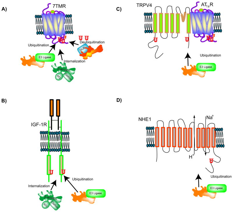
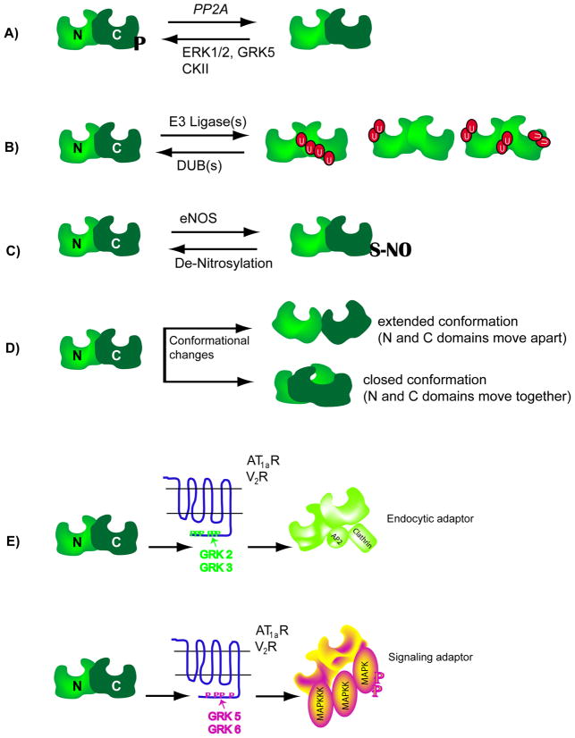
β-Arrestins function as endocytic adaptors and mediate trafficking of a variety of cell-surface receptors, including seven-transmembrane receptors (7TMRs). In the case of 7TMRs, β-arrestins carry out these tasks while simultaneously inhibiting upstream G-protein-dependent signaling and promoting alternate downstream signaling pathways. The mechanisms by which β-arrestins interact with a continuously expanding ensemble of protein partners and perform their multiple functions including trafficking and signaling are currently being uncovered. Molecular changes at the level of protein conformation as well as post-translational modifications of β-arrestins probably form the basis for their dynamic interactions during receptor trafficking and signaling. It is becoming increasingly evident that β-arrestins, originally discovered as 7TMR adaptor proteins, indeed have much broader and more versatile roles in maintaining cellular homeostasis. In this review paper, we assess the traditional and novel functions of β-arrestins and discuss the molecular attributes that might facilitate multiple interactions in regulating cell signaling and receptor trafficking.


Figures

Figure 1
Figure 1. Multifaceted functions of β-arrestins
Desensitization: Agonist-stimulation of 7TMRs leads to G protein coupling and activation, following which the receptors are rapidly phosphorylated by G protein-coupled receptor kinases (GRKs). Phosphorylated receptors present high affinity binding surfaces to recruit the cytosolic adaptors, β-arrestins. Steric binding by β-arrestin interferes with further G protein coupling leading to the desentization of G protein-dependent signaling. β-arrestins also scaffold the second messenger degrading enzymes (phosphodiesterase 4D, PDE4D that degrades cAMP and diacyl glycerol kinase or DAG-K that converts diacyl glycerol to phosphatidic acid). Endocytosis: Agonist-stimulation promotes rapid internalization of cell-surface 7TMRs into clathrin-coated vesicles. This internalization is facilitated by β-arrestin binding, which has specific binding domains for clathrin and AP2 interactions. β-arrestin binding to other endocytic proteins is also required for efficient receptor internalization: N ethyl maleimide fusion protein, NSF and ADP ribosylation factor 6, Arf6. The interaction between β-arrestin and the E3 ubiquitin ligase Mdm2 (mouse double minute 2) promotes ubiquitination of β-arrestin, that facilitates robust binding of β-arrestin with both cargo (7TMR) as well as endocytic machinery (clathrin and AP2). Receptor internalization is followed by post-endocytic sorting of internalized receptors for recycling or lysosomal degradation. Signaling: β-arrestin acquires an active conformation upon forming a complex with agonist-stimulated 7TMRs and scaffolds MAP kinase, MAP kinase kinase, and MAP kinase kinase kinase, leading to the robust activation of MAP kinase, and subsequently targets the activated kinase to distinct subcellular compartments. Such β-arrestin-dependent MAP Kinase activity has been shown to regulate cellular chemotaxis, apoptosis, cancer metastasis and protein translation.
Figure 2
Figure 2. β-arrestins function as versatile adaptors for different cell-surface receptors
A) For 7TMRs, β-arrestins act as clathrin-AP2 adaptors to facilitate receptor internalization, E3 ubiquitin ligase and deubiquitinase adaptors to facilitate receptor ubiquitination and deubiquitination. B) For the insulin-like growth factor receptor (IGF1R), β-arrestin1 functions as a clathrin adaptor as well as an E3 ligase adaptor, facilitating internalization and ubiquitination of IGF-activated receptors. C) The cell surface Transient Receptor Potential Vanilloid 4 channel (TRPV4) remains complexed with the Angiotensin 1a receptor (AT1aR) and upon AT1aR activation, becomes ubiquitinated in a β-arrestin1 dependent manner. In this process, β-arrestin1 functions as an E3 ubiquitin ligase adaptor. D) The Na+/H+ exchanger channel NHE1 is internalized and ubiquitinated in a β-arrestin-dependent manner.
Figure 3
Figure 3. Schematics showing molecular changes in β-arrestin
A) β-arrestins are phosphorylated at a seryl or threonyl residues in the C-terminal domain (see text for details) and become dephosphorylated upon 7TMR activation. Dephosphorylated β-arrestins interact more efficiently with clathrin than phosphorylated forms. Upon receptor internalization, β-arrestins become rephosphorylated. ERK1/2: extra cellular signal regulated kinase, GRK: G protein-coupled receptor kinase, CKII: casein kinase II, PP2A: protein phosphatase 2A. B) 7TMR activation leads to ubiquitination of β-arrestin2, the site of ubiquitination is unique to a particular 7TMR-β-arrestin pair, thus leading to differential modifications or differential display of ubiquitinated β-arrestins. C) The C-terminal cysteine residue in β-arrestin2 is nitrosylated and this modification promotes its interaction with clathrin. D) Upon binding to 7TMRs, the β-arrestin molecule undergoes conformational rearrangement, which could either push the amino (N) and carboxyl (C) domains apart or pull them together. The conformational changes have been assessed by various biochemical methods (limited proteolysis of purified β-arrestins, Resonance energy transfer between fluorophores attached to the two domains etc). E) When β-arrestin2 is recruited to the AT1aR or V2R that are phosphorylated by GRK2 or GRK3 isoforms, it is able to function as an endocytic adaptor, but not as a signaling adaptor. In contrast, when β-arrestin2 binds to the same receptors that are phosphorylated by GRK5 or GRK6 isoforms, it functions as a signaling adaptor leading to robust activation of MAP kinases ERK 1 and 2. It is suggested that the different GRK isoforms set respective phosphorylation ‘bar codes’ to dictate the downstream activity of β-arrestin.

References

    1. Lefkowitz RJ. Historical review: a brief history and personal retrospective of seven-transmembrane receptors. Trends Pharmacol Sci. 2004;25 (8):413–422. - PubMed
    1. Yamaki K, et al. Molecular cloning of the S-antigen cDNA from bovine retina. Biochem Biophys Res Commun. 1987;142 (3):904–910. - PubMed
    1. Lohse MJ, et al. beta-Arrestin: a protein that regulates beta-adrenergic receptor function. Science. 1990;248 (4962):1547–1550. - PubMed
    1. Attramadal H, et al. Beta-arrestin2, a novel member of the arrestin/beta-arrestin gene family. J Biol Chem. 1992;267 (25):17882–17890. - PubMed
    1. Smith WC, et al. Cloning and functional characterization of salamander rod and cone arrestins. Invest Ophthalmol Vis Sci. 2000;41 (9):2445–2455. - PubMed

Substances