. 2011 Sep;85(17):8777-88.
doi: 10.1128/JVI.02533-10. Epub 2011 Jun 15.

Peptidyl-prolyl isomerase Pin1 is a cellular factor required for hepatitis C virus propagation

Affiliations

Peptidyl-prolyl isomerase Pin1 is a cellular factor required for hepatitis C virus propagation

Yun-Sook Lim et al. J Virol. 2011 Sep.

Abstract

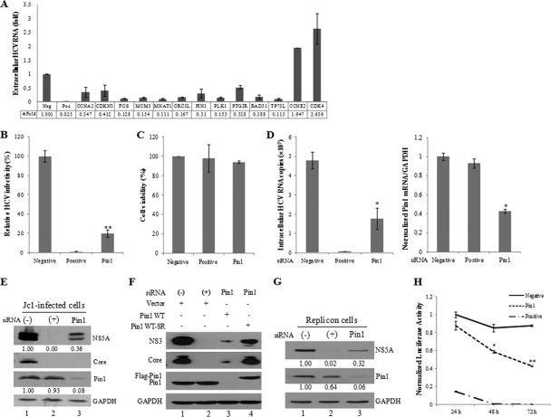
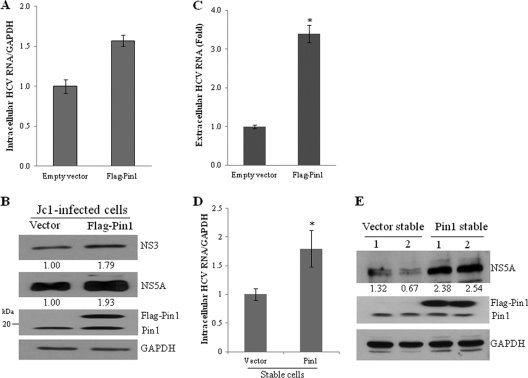
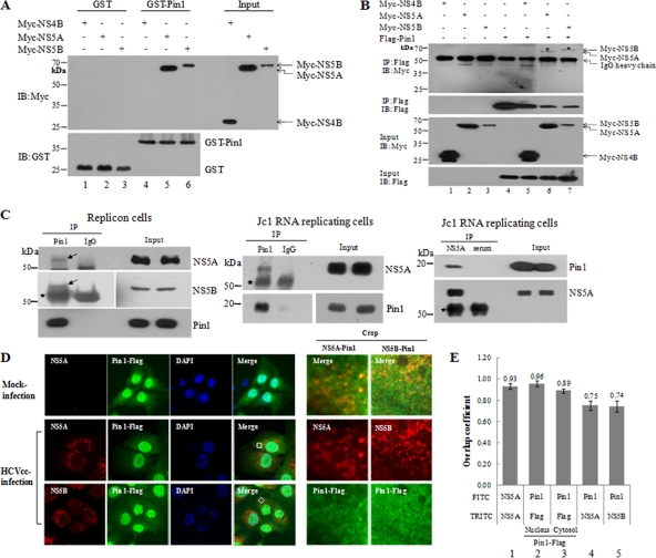
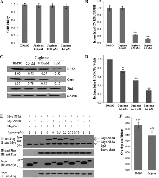
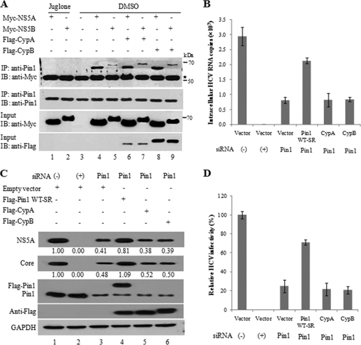
The life cycle of hepatitis C virus (HCV) is highly dependent on cellular factors. Using small interfering RNA (siRNA) library screening, we identified peptidyl-prolyl cis-trans isomerase NIMA-interacting 1 (Pin1) as a host factor involved in HCV propagation. Here we demonstrated that silencing of Pin1 expression resulted in decreases in HCV replication in both HCV replicon cells and cell culture-grown HCV (HCVcc)-infected cells, whereas overexpression of Pin1 increased HCV replication. Pin1 interacted with both the NS5A and NS5B proteins. However, Pin1 expression was increased only by the NS5B protein. Both the protein binding and isomerase activities of Pin1 were required for HCV replication. Juglone, a natural inhibitor of Pin1, inhibited HCV propagation by inhibiting the interplay between the Pin1 and HCV NS5A/NS5B proteins. These data indicate that Pin1 modulates HCV propagation and may contribute to HCV-induced liver pathogenesis.


Figures

Fig. 1.
Fig. 1.
Pin1 is essential for HCV replication. (A) Huh7.5 cells were transfected with 20 nM siRNAs for 48 h and were then infected with HCV Jc1 for 4 h. At 2 days postinfection, culture supernatants were collected, and extracellular HCV RNAs were quantified by qPCR. From the siRNA library targeting 131 human cell cycle genes, 13 siRNAs that altered (increased or decreased) the level of HCV production 2-fold from that for the negative control were selected. (B) Huh7.5 cells were transfected with 20 nM negative (−), positive (+), or Pin1 siRNA duplexes for 48 h, followed by infection with Jc1 for 4 h. At 2 days postinfection, HCV infectivity was determined by a focus-forming assay. (C) Huh7.5 cells were transfected with the indicated siRNAs. At 96 h after transfection, cell viability was assessed by an MTT assay. (D) Both intracellular RNA (left) and Pin1 mRNA (right) isolated from cells treated as described in the legend to panel B were analyzed by qPCR. (E) Total-cell lysates harvested from cells treated as described in the legend to panel B were immunoblotted with the indicated antibodies. (F) Huh7.5 cells were transfected with the indicated siRNA. At 2 days after siRNA transfection, cells were infected with Jc1 for 4 h and were then transfected with 3 μg of a vector or plasmid expressing wild-type Pin1 (Flag-Pin1 WT) or siRNA-resistant Pin1 (Flag-Pin1 WT-SR). Protein expression was determined by immunoblot analysis at 2 days after transfection. (−), the universal negative-control siRNA; (+), siRNA targeting the 5′ NTR of Jc1 as a positive control. (G) Subgenomic replicon cells were transfected with the indicated siRNAs for 96 h. Protein expression was determined by immunoblot analysis. The band intensity was quantified using ImageJ software. (H) Huh7.5 cells were transfected with in vitro-transcribed HCV-Luc RNA. At 2 days after transfection, cells were transfected with a negative-control, positive-control, or Pin1 siRNA. Luciferase activity was measured at the indicated times. Asterisks indicate significant differences (*, P < 0.05; **, P < 0.01) from activity for the negative control.
Fig. 2.
Fig. 2.
Overexpression of Pin1 increases HCV propagation. (A) Huh7.5 cells were infected with Jc1 for 48 h and were then transiently transfected with 2 μg of either a vector or the Flag-Pin1 plasmid. At 48 h after transfection, intracellular HCV RNAs were analyzed by qPCR. (B) Total-cell lysates harvested from these cells were immunoblotted with the indicated antibodies. (C) Extracellular RNAs isolated from the culture supernatant were quantified by qPCR. Experiments were carried out in duplicate. Error bars indicate standard deviations. (D) Vector-stable and Pin1-stable cells were infected with Jc1. At 48 h postinfection, intracellular HCV RNAs were quantified by qPCR. The asterisk indicates a significant difference (*, P < 0.05) from the value for the vector control. (E) Two clones each of vector-stable and Pin1-stable cells were infected with Jc1. At 48 h postinfection, total-cell lysates were immunoblotted with the indicated antibodies. The band intensity was quantified using ImageJ software.
Fig. 3.
Fig. 3.
Pin1 interacts directly with both the HCV NS5A and NS5B proteins. (A) HEK293T cells were transfected with Myc-NS4B, Myc-NS5A, or Myc-NS5B. At 36 h after transfection, total-cell lysates were harvested and were incubated with either purified GST or GST-Pin1. (Top) Complexes were precipitated with glutathione beads, and bound proteins were analyzed by immunoblotting (IB) using an anti-Myc antibody. (Bottom) The expression of the GST and GST-Pin1 proteins was confirmed with an anti-GST antibody. (B) HEK293T cells were transfected with the indicated combinations of expression plasmids. (Top panel) At 36 h after transfection, cell lysates were immunoprecipitated (IP) with an anti-Flag monoclonal antibody, and bound proteins were then detected by IB analysis. (Lower panels) Protein expression of Myc-NS4B, Myc-NS5A, Myc-NS5B, and Flag-Pin1 in the same cell lysates was also verified by immunoblot analysis. (C) (Left) Total-cell lysates harvested from HCV replicon cells were immunoprecipitated with either an anti-Pin1 antibody or IgG. Bound protein was detected by immunoblotting with an antibody against either NS5A or NS5B. (Center) Huh7.5 cells were electroporated with 10 μg Jc1 RNA and were incubated for 3 days. Cell lysates were immunoprecipitated with either an anti-Pin1 antibody or IgG. Bound protein was immunoblotted with an anti-NS5A antibody. (Right) Reciprocally, the same cell lysates were immunoprecipitated with either a rabbit anti-NS5A antibody or a rabbit control serum. Bound protein was immunoblotted with an anti-Pin1 antibody. The asterisk indicates a heavy chain. (D) Huh7.5 cells were infected with HCV Jc1 for 4 h. At 2 days postinfection, cells were transfected with a plasmid expressing Flag-tagged Pin1. At 24 h after transfection, cells were fixed in 4% paraformaldehyde, and immunofluorescence staining was performed by using an anti-Pin1 monoclonal antibody and fluorescein isothiocyanate-conjugated goat anti-mouse IgG to detect Pin1 (green) and a rabbit anti-NS5A or anti-NS5B antibody and TRITC-conjugated goat anti-rabbit IgG to detect NS5A (red) or NS5B (red). Dual staining showed colocalization of Pin1 and NS5A as yellow fluorescence in the merged image. Cells were counterstained with 4′,6-diamidino-2-phenylindole (DAPI) to label nuclei. The boxed area in the merged image is enlarged on the right (crop). (E) Huh7.5 cells infected with HCV Jc1 were fixed, incubated with a rabbit anti-NS5A antibody, and further incubated with either FITC- or TRITC-conjugated anti-rabbit antibodies (bar 1). Flag-Pin1-stable cells were fixed, incubated with both a mouse anti-Pin1 and a rabbit anti-Flag antibody, and further incubated with both TRITC-conjugated anti-mouse and FITC-conjugated anti-rabbit antibodies (bars 2 and 3). Colocalization of NS5A (bar 4) and NS5B (bar 5) with Pin1 was quantified by Manders' overlap coefficient as described in Materials and Methods.
Fig. 4.
Fig. 4.
NS5B but not NS5A increases Pin1 expression. (A) (Left) Huh7.5 cells were either mock infected or infected with HCV Jc1 for 4 h. Total-cell lysates harvested at 4 days postinfection were immunoblotted with the indicated antibodies. (Center) Blot images were scanned, and protein pixel intensity was measured using ImageJ software. (Right) Total RNAs were extracted, and qPCR was performed to quantify the Pin1 mRNA level. Data from two independent experiments were quantified. The asterisk indicates a significant difference (P < 0.05) between mock-infected and HCV Jc1-infected cells. (B) (Left) Equal amounts of cell lysates harvested from either IFN-cured or subgenomic replicon cells were immunoblotted with the indicated antibodies. (Center) Blot images were scanned, and the protein pixel intensity was measured using ImageJ software. (Right) Total RNAs were extracted, and qPCR was performed to quantify the Pin1 mRNA level. Data from two independent experiments were quantified. Asterisks indicate a significant difference (P < 0.05) between IFN-cured and replicon cells. (C) Huh7 cells were transfected with either a vector, Myc-tagged NS5A, or Myc-tagged NS5B. At 48 h after transfection, protein expression was determined by immunoblot analysis.
Fig. 5.
Fig. 5.
Both the binding and isomerase activities of Pin1 are essential for HCV replication. (A) Huh7.5 cells were transfected with the indicated combinations of expression plasmids. (Top panel) At 48 h after transfection, cell lysates were immunoprecipitated (IP) with an anti-Flag monoclonal antibody, and bound proteins were then detected by an immunoblot (IB) assay. (Lower panels) Protein expression in the same cell lysates was verified with the indicated antibodies. Pin1 WT-SR, siRNA-resistant mutant Pin1; Pin1 S16A-SR, siRNA-resistant binding-defective mutant Pin1; Pin1 C113A-SR, siRNA-resistant isomerase-inactive mutant Pin1. The asterisk indicates the IgG heavy chain. (B) Huh7.5 cells were transfected with the indicated siRNA. At 2 days after siRNA transfection, cells were infected with Jc1 for 4 h and were then transfected with 2 μg of a plasmid expressing wild-type Pin1 (Pin1 WT), Pin1 S16A-SR, Pin1 C113A-SR, or Pin1 WT-SR. Intracellular RNAs isolated at 2 days after Pin1 transfection were analyzed by qPCR. (C) Protein expression levels of both HCV and host cells were determined by immunoblot analysis using the indicated antibodies. (D) The effects of Pin1 mutants on HCV infectivity were determined by focus-forming assays. Experiments were carried out in duplicate. Error bars indicate standard deviations.
Fig. 6.
Fig. 6.
Juglone represses HCV replication by inhibiting the interplay between Pin1 and HCV proteins. (A) Huh7.5 cells were infected with Jc1. One day after HCV infection, cells were treated with different concentrations of juglone for 1 day. Cell viability was assessed by the MTT assay. (B) Intracellular RNAs isolated from these cells were analyzed by qPCR. (C) Protein expression in these cells was analyzed by immunoblotting (IB). (D) Extracellular RNAs isolated from these cells were quantified by qPCR. Experiments were carried out in duplicate. Asterisks indicate significant differences (*, P < 0.05; **, P < 0.01) from results with DMSO (vehicle). (E) HEK293T cells were transfected with the indicated combinations of expression plasmids. (Top panel) At 24 h after transfection, cell lysates were immunoprecipitated (IP) with an anti-Flag monoclonal antibody, and bound proteins were then detected by immunoblot analysis using an anti-Myc antibody. (Lower panels) The expression of the NS5A, NS5B, and Pin1 proteins in the same cell lysates was confirmed by immunoblot analysis using the indicated antibodies. (F) Huh7.5 cells infected with HCV Jc1 were transiently transfected with a plasmid expressing Flag-tagged Pin1 at 24 h postinfection. Cells were then treated with either DMSO (vehicle) or 1 μM of juglone at 24 h after transfection. Samples were fixed and were incubated with both anti-NS5A and anti-Flag antibodies. The overlap coefficient was determined as described in Materials and Methods using immunofluorescence data. *, P < 0.05.
Fig. 7.
Fig. 7.
Pin1, CypA, and CypB are distinctively involved in HCV replication. (A) HEK293T cells were transfected with the indicated combinations of expression plasmids. At 24 h after transfection, cells were treated with either 1 μM juglone or a vehicle (DMSO) for 24 h. (Top panel) Cell lysates were immunoprecipitated (IP) with an anti-Pin1 polyclonal antibody, and bound proteins were then detected by immunoblot (IB) analysis. (Lower panels) The expression of the NS5A, NS5B, Pin1, CypA, and CypB proteins in the same cell lysates was confirmed by immunoblot analysis using the indicated antibodies. (B) Huh7.5 cells were transfected with the indicated siRNA. At 2 days after siRNA transfection, cells were infected with Jc1 for 4 h and were then transfected with 2 μg of an empty vector or a plasmid expressing WT-SR, CypA, or CypB. Intracellular RNAs were isolated and were analyzed by qPCR. (C) Both viral and cellular proteins isolated from these cells were analyzed by immunoblot analysis using the indicated antibodies. The band intensity was quantified as described for Fig. 1. (D) Virus titers were determined by focus-forming assays. Experiments were carried out in duplicate.

Similar articles

Cited by

References

    1. Chao S. H., Greenleaf A. L., Price D. H. 2001. Juglone, an inhibitor of the peptidyl-prolyl isomerase Pin1, also directly blocks transcription. Nucleic Acids Res. 29:767–773 - PMC - PubMed
    1. Chatterji U., et al. 2010. HCV resistance to cyclosporin A does not correlate with a resistance of the NS5A-cyclophilin A interaction to cyclophilin inhibitors. J. Hepatol. 53:50–56 - PMC - PubMed
    1. Choi S. H., Hwang S. B. 2006. Modulation of the transforming growth factor-beta signal transduction pathway by hepatitis C virus nonstructural 5A protein. J. Biol. Chem. 281:7468–7478 - PubMed
    1. Choi S. H., Jeong S. H., Hwang S. B. 2007. Large hepatitis delta antigen modulates transforming growth factor-beta signaling cascades: implication of hepatitis delta virus-induced liver fibrosis. Gastroenterology 132:343–357 - PubMed
    1. Fernandes F., Ansari I. U., Striker R. 2010. Cyclosporine inhibits a direct interaction between cyclophilins and hepatitis C NS5A. PLoS One 5:e9815. - PMC - PubMed

Publication types

MeSH terms

Substances

LinkOut - more resources