. 2011 Aug;85(16):8208-16.
doi: 10.1128/JVI.00114-11. Epub 2011 Jun 15.

E6 and E7 from human papillomavirus type 16 cooperate to target the PDZ protein Na/H exchange regulatory factor 1

Affiliations

E6 and E7 from human papillomavirus type 16 cooperate to target the PDZ protein Na/H exchange regulatory factor 1

Rosita Accardi et al. J Virol. 2011 Aug.

Abstract

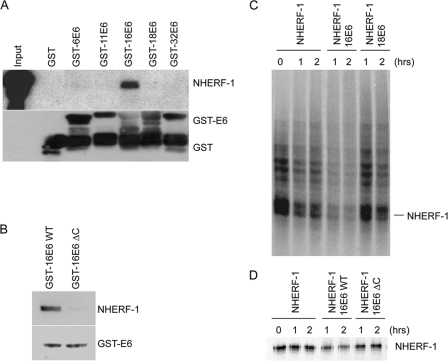
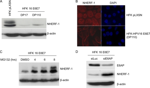
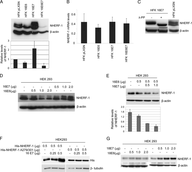
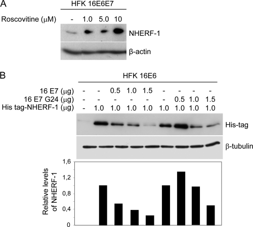
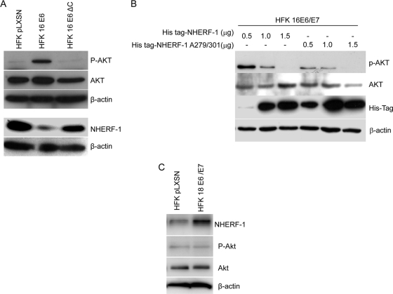
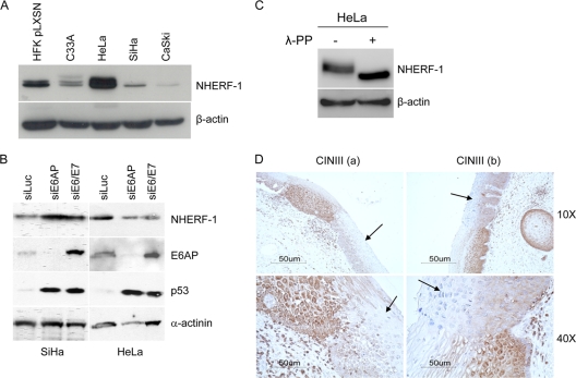
Previous studies have shown that the PDZ-binding motif of the E6 oncoprotein from the mucosal high-risk (HR) human papillomavirus (HPV) types plays a key role in HPV-mediated cellular transformation in in vitro and in vivo experimental models. HR HPV E6 oncoproteins have the ability to efficiently degrade members of the PDZ motif-containing membrane-associated guanylate kinase (MAGUK) family; however, it is possible that other PDZ proteins are also targeted by E6. Here, we describe a novel interaction of HPV type 16 (HPV16) E6 with a PDZ protein, Na(+)/H(+) exchange regulatory factor 1 (NHERF-1), which is involved in a number of cellular processes, including signaling and transformation. HPV16 E6 associates with and promotes the degradation of NHERF-1, and this property is dependent on the C-terminal PDZ-binding motif of E6. Interestingly, HPV16 E7, via the activation of the cyclin-dependent kinase complexes, promoted the accumulation of a phosphorylated form of NHERF-1, which is preferentially targeted by E6. Thus, both oncoproteins appear to cooperate in targeting NHERF-1. Notably, HPV18 E6 is not able to induce NHERF-1 degradation, indicating that this property is not shared with E6 from all HR HPV types. Downregulation of NHERF-1 protein levels was also observed in HPV16-positive cervical cancer-derived cell lines, such as SiHa and CaSki, as well as HPV16-positive cervical intraepithelial neoplasia (CIN). Finally, our data show that HPV16-mediated NHERF-1 degradation correlates with the activation of the phosphatidylinositol-3'-OH kinase (PI3K)/AKT signaling pathway, which is known to play a key role in carcinogenesis.


Figures

Fig. 1.
Fig. 1.
HPV16 E6 binds NHERF-1 and promotes its degradation in in vitro assays. (A and B) C33A cell extracts were incubated with the different GST fusion proteins indicated and loaded onto SDS-PAGE gel. The amounts of NHERF-1 associated with the different GST E6 proteins were determined by immunoblotting using a specific anti-NHERF-1 antibody. One-tenth of the total cellular extract (60 μg) used in the GST pulldown assay (input) was used for SDS-PAGE. (C and D) In vitro-translated NHERF-1 was incubated at 30°C for 0, 1, or 2 h either alone or with in vitro-translated HPV16 or HPV18 E6 (C) or wild-type (WT) or ΔC mutant HPV16 E6 (D). The remaining targeted proteins were visualized by SDS-PAGE and autoradiography.
Fig. 2.
Fig. 2.
NHERF-1 protein levels are downregulated in HFKs expressing HPV16 E6 and E7. (A) Protein extracts of primary keratinocytes transduced with empty retrovirus vector (HFK pLXSN) or keratinocytes expressing HPV16 E6 and E7 genes (HFK 16 E6E7) were analyzed by immunoblotting using anti-NHERF-1 and anti-β-actin antibodies. β-Actin staining was included as a loading control. (B) Primary HFKs transduced with empty retrovirus (pLXSN) and HFKs expressing HPV16 E6 and E7 were seeded on coverslips. After immunofluorescent staining for NHERF-1 with specific antibodies, fluorescent signal was visualized using fluorescence microscopy. (C) HFKs expressing HPV16 E6 and E7 were treated with the protease inhibitor MG132 for the times indicated. NHERF-1 protein levels were determined by SDS-PAGE and immunoblotting. (D) HFKs expressing HPV16 E6 and E7 were transiently transfected with the indicated small interfering RNAs. After preparation of total protein extracts, NHERF-1, E6AP, and β-actin protein levels were determined by immunoblotting using specific antibodies.
Fig. 3.
Fig. 3.
HPV16 E6 and E7 cooperate in NHERF-1 degradation. (A) Protein extracts of primary keratinocytes transduced with empty retrovirus vector (HFK pLXSN) or recombinant retroviruses, as indicated, were analyzed by immunoblotting using anti-NHERF-1 and anti-β-actin antibodies (top). The intensities of the protein bands in three independent experiments were quantified (bottom). (B) Total RNA was also extracted from the indicated cells, and NHERF-1 or GAPDH mRNA levels were measured by quantitative RT-PCR. y axis numbers represent arbitrary units of NHERF-1 mRNA levels in indicated cells standardized to the GAPDH levels. The data are the means of three independent experiments. The differences between the NHERF-1 levels in the different cells are not statically significant. (C) Protein extracts from HFKs transduced with empty (pLXSN) or HPV16 E7 retrovirus were incubated in the presence and absence of λ-PP and analyzed by immunoblotting using anti-NHERF-1 and anti-β-actin antibodies. (D) HEK293 cells were transfected with increasing concentrations of pLXSN HPV16 E6 or E7 as indicated. After 24 h, protein extracts were analyzed by immunoblotting. (E) HEK293 cells were transfected with pLXSN expressing HPV16 E6 together with increasing concentrations of pLXSN expressing HPV16 E7 as indicated. After 24 h, protein extracts were analyzed by immunoblotting (top). The intensities of the bands were quantified in three independent experiments and are represented in the histogram (bottom). (F) HEK293 cells were transfected with pcDNA3 expressing His-tagged NHERF-1 (His-NHERF-1), wild type or A279/A301 mutant, together with increasing concentrations of pLSXN expressing HPV16 E7 as indicated. After 24 h, protein extracts were analyzed by immunoblotting. (G) HEK293 cells were transfected with increasing concentrations of pLXSN expressing HPV18 E6 or E7 as indicated. After 24 h, protein extracts were analyzed by immunoblotting.
Fig. 4.
Fig. 4.
CDK-mediated NHERF-1 phosphorylation is required for its degradation induced by HPV16 E6. (A) HFKs expressing HPV16 E6 and E7 were treated with the CDK inhibitor roscovitine at the indicated concentrations, and NHERF-1 protein levels were determined by SDS-PAGE and immunoblotting. (B) HFKs expressing HPV16 E6 were transfected with pLXSN expressing His-NHERF-1, together with increasing concentrations of pLSXN expressing HPV16 E7, wild type or the glycine 24 mutant (G24), as indicated. After 24 h, protein extracts were analyzed by immunoblotting (top), and the intensities of the protein bands were quantified (bottom).
Fig. 5.
Fig. 5.
HPV16 E6-induced NHERF-1 degradation leads to activation of AKT signaling. (A) Protein extracts of primary keratinocytes transduced with empty retrovirus vector (pLXSN), or HPV16 E6/E7-expressing retrovirus were analyzed by immunoblotting using anti-NHERF-1, anti-AKT, anti-phospho-AKT, and anti-β-actin antibodies. (B) HFKs expressing HPV16 E6 and E7 were transfected with increasing concentrations of pcDNA3 expressing His-NHERF-1, wild type or A279/A301 mutant. After 24 h, protein extracts were analyzed by immunoblotting. (C) Protein extracts of primary keratinocytes transduced with empty retrovirus vector (pLXSN) or HPV18 E6/E7-expressing retrovirus were analyzed by immunoblotting using anti-NHERF-1, anti-AKT, anti-phospho-AKT, and anti-β-actin antibodies.
Fig. 6.
Fig. 6.
NHERF-1 levels are downregulated in HPV16-positive cervical cancer cell lines and in cervical lesions. (A) Protein extracts of different cell lines as indicated were prepared and analyzed by immunoblotting using anti-NHERF-1 and anti-β-actin antibodies. (B) SiHa and HeLa cells were transiently transfected with the indicated small interfering RNAs. After preparation of total protein extracts, NHERF-1, E6AP, p53, and α-actinin protein levels were determined by immunoblotting using specific antibodies. (C) Protein extracts from HeLa cells were incubated in the presence and absence of λ-PP and analyzed by immunoblotting using anti-NHERF-1 and anti-β-actin antibodies. (D) Sections of HPV16-positive cervical intraepithelial neoplasia grade III (CINIII) from two different women (a and b) were stained with a specific anti-NHERF-1 antibody. Arrows indicate the dysplastic regions. The images are shown at two magnifications (×10 and ×40).

References

    1. Accardi R., et al. 2006. Skin human papillomavirus type 38 alters p53 functions by accumulation of deltaNp73. EMBO Rep. 7:334–340 - PMC - PubMed
    1. Bretscher A., Edwards K., Fehon R. G. 2002. ERM proteins and merlin: integrators at the cell cortex. Nat. Rev. Mol. Cell Biol. 3:586–599 - PubMed
    1. Caldeira S., et al. 2003. The E6 and E7 proteins of cutaneous human papillomavirus type 38 display transforming properties. J. Virol. 77:2195–2206 - PMC - PubMed
    1. Georgescu M. M., Morales F. C., Molina J. R., Hayashi Y. 2008. Roles of NHERF1/EBP50 in cancer. Curr. Mol. Med. 8:459–468 - PubMed
    1. Ghittoni R., et al. 2010. The biological properties of E6 and E7 oncoproteins from human papillomaviruses. Virus Genes 40:1–13 - PubMed

Publication types

MeSH terms

Substances

LinkOut - more resources