. 2011 Aug;85(16):8285-92.
doi: 10.1128/JVI.00741-11. Epub 2011 Jun 15.

Nuclear envelope breakdown can substitute for primary envelopment-mediated nuclear egress of herpesviruses

Affiliations

Nuclear envelope breakdown can substitute for primary envelopment-mediated nuclear egress of herpesviruses

Barbara G Klupp et al. J Virol. 2011 Aug.

Abstract

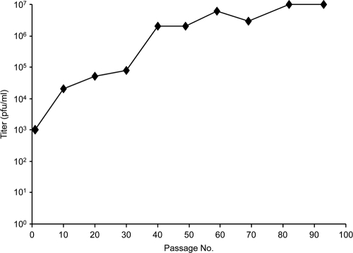
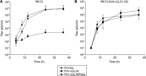
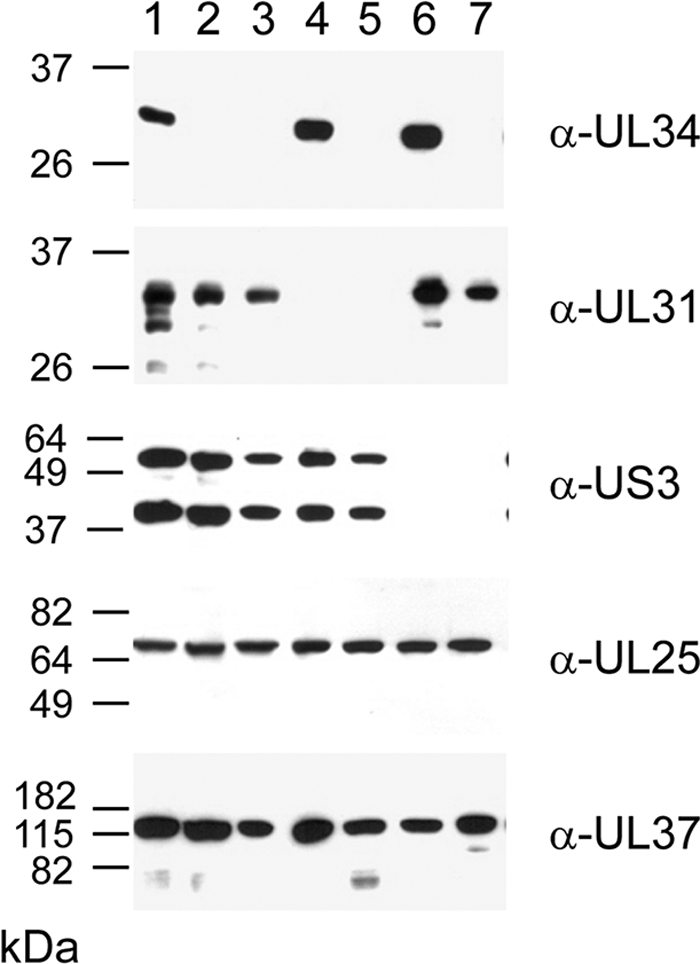
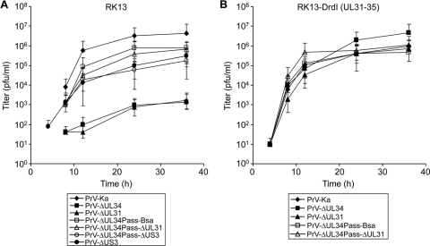
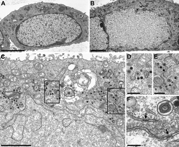
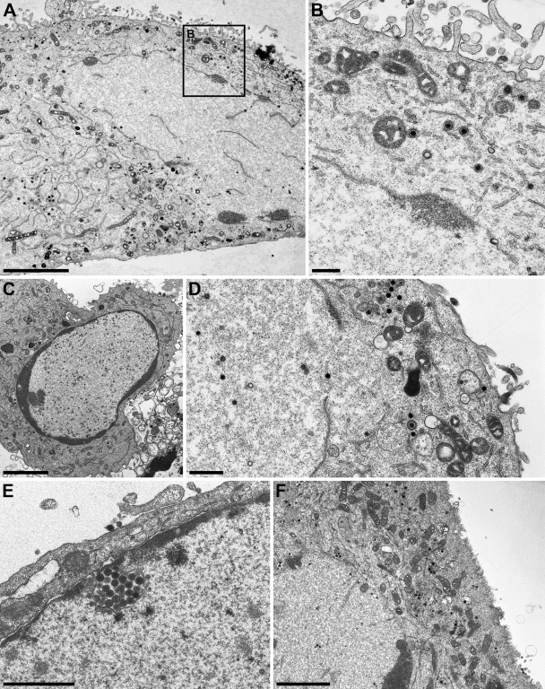
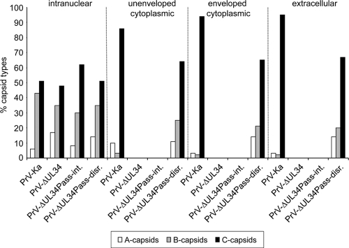
Herpesvirus nucleocapsids assemble in the nucleus but mature to infectious virions in the cytoplasm. To gain access to this cellular compartment, nucleocapsids are translocated to the cytoplasm by primary envelopment at the inner nuclear membrane and subsequent fusion of the primary envelope with the outer nuclear membrane. The conserved viral pUL34 and pUL31 proteins play a crucial role in this process. In their absence, viral replication is strongly impaired but not totally abolished. We used the residual infectivity of a pUL34-deleted mutant of the alphaherpesvirus pseudorabies virus (PrV) for reversion analysis. To this end, PrV-ΔUL34 was serially passaged in rabbit kidney cells until final titers of the mutant virus PrV-ΔUL34Pass were comparable to those of wild-type PrV. PrV-ΔUL34Pass produced infectious progeny independently of the pUL34/pUL31 nuclear egress complex and the pUS3 protein kinase. Ultrastructural analyses demonstrated that this effect was due to virus-induced disintegration of the nuclear envelope, thereby releasing immature and mature capsids into the cytosol for secondary envelopment. Our data indicate that nuclear egress primarily serves to transfer capsids through the intact nuclear envelope. Immature and mature intranuclear capsids are competent for further virion maturation once they reach the cytoplasm. However, nuclear egress exhibits a strong bias for nucleocapsids, thereby also functioning as a quality control checkpoint which is abolished by herpesvirus-induced nuclear envelope breakdown.


Figures

Fig. 1.
Fig. 1.
Diagram of mutant viruses. The PrV genome consisting of long (UL) and short (US) unique regions and inverted repeat sequences (IR and TR), as well as location and numbering of BamHI restriction fragments, is shown. Locations of the genomic regions shown enlarged are indicated. (A) UL34 gene region with relevant restriction sites. Open reading frames are drawn as rectangles. The regions which had been deleted for generation of PrV-ΔUL34 (SalI/SphI) and PrV-ΔUL34Pass-Bsa (SalI/BsaBI) are delineated by dashed lines. Insertion of the reporter gene in PrV-ΔUL34 is indicated. (B) UL31 gene region and relevant restriction sites. Locations of primers used for amplification of UL31 flanking regions are indicated by bent arrows. Deletion of UL31 coding sequences is shown by dashed lines, and insertion of the GFP expression cassette is indicated. (C) Substitution of US3 coding sequences between the dashed lines with a GFP reporter cassette is indicated (10).
Fig. 2.
Fig. 2.
Selection of PrV-ΔUL34Pass. Rabbit kidney (RK13) cells were infected with PrV-ΔUL34 and passaged by initial repeated trypsinization and reseeding of infected cells followed by passaging of supernatants after a complete cytopathic effect had developed. Extracellular infectivity was determined on RK13 cells. After ca. 90 passages, virus progeny was plaque purified and further analyzed.
Fig. 3.
Fig. 3.
One-step growth kinetics of wild-type and mutant viruses. RK13 (A) or RK13-DrdI (B) cells were infected with PrV-Ka, PrV-ΔUL34, or PrV-ΔUL34Pass at an MOI of 5 and analyzed at the indicated times after low-pH treatment. The cells were lysed by freezing and thawing, and progeny virus titers were determined on RK13-DrdI cells by plaque assays. Mean values of at least two independent experiments were calculated, and corresponding minimum and maximum values were plotted.
Fig. 4.
Fig. 4.
Immunoblots of infected cells. RK13 cells infected at an MOI of 5 with PrV-Ka (lane 1), PrV-ΔUL34 (lane 2), PrV-ΔUL34Pass (lane 3), PrV-ΔUL31 (lane 4), PrV-ΔUL34Pass-ΔUL31 (lane 5), PrV-ΔUS3 (lane 6), or PrV-ΔUL34Pass-ΔUS3 (lane 7) were lysed 18 h postinfection, and proteins were separated on SDS-10% or 12% polyacrylamide gels. Parallel blots were incubated with rabbit sera raised against pUL34, pUL31, pUS3, pUL25, or pUL37. The locations of molecular mass markers are indicated on the left.
Fig. 5.
Fig. 5.
Growth kinetics of PrV-ΔUL34Pass-Bsa mutants lacking UL31 or US3. RK13 (A) or RK13-DrdI (B) cells were infected with PrV-Ka, PrV-ΔUL34, PrV-ΔUL31, PrV-ΔUL34Pass-Bsa, PrV-ΔUL34Pass-ΔUL31, PrV-ΔUS3, or PrV-ΔUL34Pass-ΔUS3 and processed as described in the legend to Fig. 3.
Fig. 6.
Fig. 6.
Ultrastructural analysis of PrV-ΔUL34Pass-infected cells. RK13 cells infected with PrV-ΔUL34 (A) or PrV-ΔUL34Pass (B to F) at an MOI of 1 were analyzed 14 h after infection by transmission electron microscopy. (A) An intact nucleus with trapped nucleocapsids is shown. (B) A partially dissolved nucleus. (C to E) Remnants of the nuclear envelope are visible together with capsids in the cytoplasm (C), several of them undergoing envelopment, magnified in panels D and E. (F) Morphologically intact nuclear pores in nuclear envelope fragments are labeled by arrows. Bars, 5 μm (A and B), 2 μm (C), 500 nm (D and E), and 200 nm (F).
Fig. 7.
Fig. 7.
Ultrastructural analysis of cells infected with PrV mutants. RK13-UL34 cells infected with PrV-ΔUL34Pass (A and B), as well as RK13 cells infected with PrV-ΔUL31 (C), PrV-ΔUL34Pass-ΔUL31 (D), PrV-ΔUS3 (E), or PrV-ΔUL34Pass-ΔUS3 (F), at an MOI of 1 were analyzed 14 h after infection by transmission electron microscopy. Bars, 5 μm (A), 500 nm (B and D), 3.7 μm (C), 1.5 μm (E), and 3 μm (F).
Fig. 8.
Fig. 8.
Quantitation of different capsid forms. Electron microscopic images of RK13 cells infected with PrV-Ka, PrV-ΔUL34, and PrV-ΔUL34Pass were used to count different capsid forms (empty A capsids, scaffold-containing B capsids, and DNA-containing C capsids) in the nucleus and nonenveloped and enveloped particles in the cytoplasm as well as in the extracellular space. Capsids in 10 cells per assay were counted. The total numbers of capsids counted were 666 for cells infected with PrV-Ka, 954 for PrV-ΔUL34-infected cells, 580 for PrV-ΔUL34Pass-infected cells with intact nuclei (PrV-ΔUL34Pass-int.; all intranuclear), and 1,188 for PrV-ΔUL34Pass-infected cells with disrupted nuclei (PrV-ΔUL34Pass-disr.).

References

    1. Bubeck A., et al. 2004. Comprehensive mutational analysis of a herpesvirus gene in the viral genome context reveals a region essential for virus replication. J. Virol. 78:8026–8035 - PMC - PubMed
    1. Farina A., et al. 2005. BFRF1 of Epstein-Barr virus is essential for efficient primary viral envelopment and egress. J. Virol. 79:3703–3712 - PMC - PubMed
    1. Farnsworth A., et al. 2007. Herpes simplex virus glycoproteins gB and gH function in fusion between the virion envelope and the outer nuclear membrane. Proc. Natl. Acad. Sci. U. S. A. 104:10187–10192 - PMC - PubMed
    1. Fuchs W., Klupp B. G., Granzow H., Osterrieder N., Mettenleiter T. C. 2002. The interacting UL31 and UL34 gene products of pseudorabies virus are involved in egress from the host-cell nucleus and represent components of primary enveloped but not mature virions. J. Virol. 76:364–378 - PMC - PubMed
    1. Granato M., et al. 2008. Deletion of Epstein-Barr virus BFLF2 leads to impaired viral DNA packaging and primary egress as well as to the production of defective viral particles. J. Virol. 82:4042–4051 - PMC - PubMed

Publication types

MeSH terms

Substances

LinkOut - more resources