

Review
doi: 10.3174/ajnr.A2404.
Epub 2011 Jun 16.
Pseudotumor cerebri: brief review of clinical syndrome and imaging findings
Affiliations
- PMID: 21680652
- PMCID: PMC7964411
- DOI: 10.3174/ajnr.A2404
Item in Clipboard
Review
Pseudotumor cerebri: brief review of clinical syndrome and imaging findings
AJNR Am J Neuroradiol.
2011 Dec.
Abstract
PTC is a clinical entity of uncertain etiology characterized by intracranial hypertension. The syndrome classically manifests with headaches and visual changes in women with obesity. Traditionally, imaging ruled out secondary causes of elevated CSF pressure but now may reveal findings frequently seen in patients with PTC, including the following: flattening of the globe, an empty sella, an enlarged ONS, protrusion and enhancement of the optic nerve head, and increased tortuosity of the optic nerve. Novel imaging methods, including MR venography, have additionally identified sinovenous stenosis as a potential indicator of PTC.
Figures
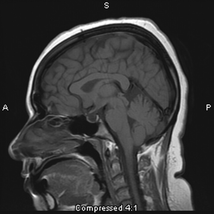
This 31-year-old woman presenting with headache is found to have an empty sella on sagittal T1-weighted MR imaging.
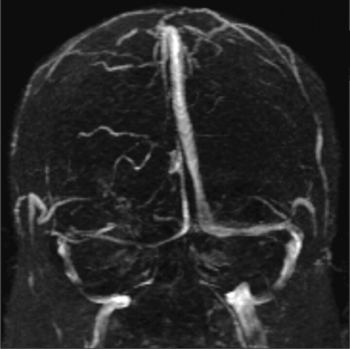
A narrowed right transverse sinus is noted in this 32-year-old woman, seen on MR venography, in addition to ONS enlargement and a partially empty sella on axial MR imaging.
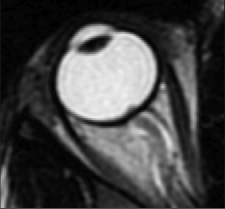
Protrusion of the right optic nerve head and horizontal tortuosity of the optic nerve are seen in this 21-year-old woman on axial T2-weighted MR imaging. Clinically, the patient presented with headaches, vision changes, and papilledema noted on examination.
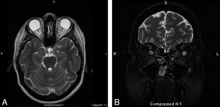
A, The ONS is widened with expanded CSF hyperintensity surrounding the optic nerve, seen on axial T2-weighted MR imaging in conjunction with posterior flattening of the globes. ONS widening is thought to coincide with papilledema, which is seen in this 27-year-old woman who presented with headaches. B, Coronal T2-weighted MR imaging in a 55-year-old woman with headache demonstrates increased peri-ONS space marked by hyperintense signal intensity surrounding the optic nerve.
References
-
- Pearce JM. From pseudotumour cerebri to idiopathic intracranial hypertension. Pract Neurol 2009;9:353–56 - PubMed
-
- Johnston I. The historical development of the pseudotumor concept. Neurosurg Focus 2001;11:1–9 - PubMed
-
- Giuseffi V, Wall M, Siegel PZ, et al. Symptoms and disease associations in idiopathic intracranial hypertension (pseudotumor cerebri): a case-control study. Neurology 1991;41:239–44 - PubMed
-
- Bandyopadhyay S, Jacobson DM. Clinical features of late-onset pseudotumor cerebri fulfilling the modified Dandy criteria. J Neuroophthalmol 2002;22:9–11 - PubMed
-
- Sylaja PN, Ahsan Moosa NV, Radhakrishnan K, et al. Differential diagnosis of patients with intracranial sinus venous thrombosis-related isolated intracranial hypertension from those with idiopathic intracranial hypertension. J Neurol Sci 2003;215:9–12 - PubMed
Publication types
MeSH terms
LinkOut - more resources
Full Text Sources
Medical