. 2011 Sep;32(8):1459-63.
doi: 10.3174/ajnr.A2517. Epub 2011 Jun 16.

Joubert syndrome and related disorders: spectrum of neuroimaging findings in 75 patients

Affiliations

Joubert syndrome and related disorders: spectrum of neuroimaging findings in 75 patients

A Poretti et al. AJNR Am J Neuroradiol. 2011 Sep.

Abstract

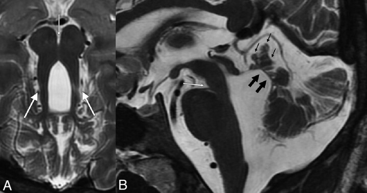
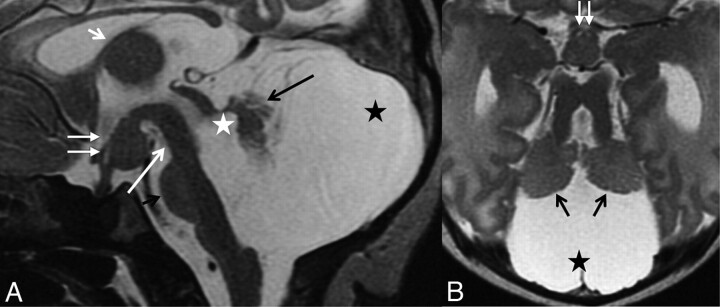
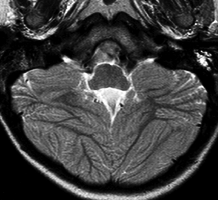
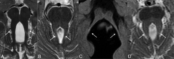
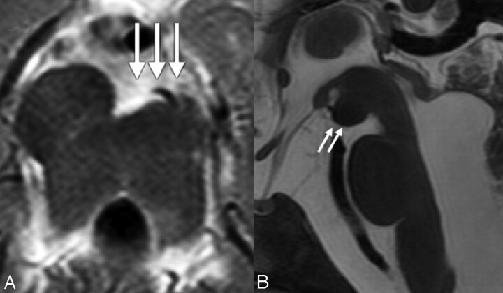
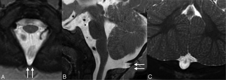
VH and MTS are the neuroimaging hallmarks of JSRD. We aimed to look at the full spectrum of neuroimaging findings in JSRD and reviewed the MR imaging of 75 patients with JSRD, including 13 siblings and 4 patients with OFD VI. All patients had VH and enlargement of the fourth ventricle. The degree of VH and the form of the MTS were variable. In most patients, the cerebellar hemispheres were normal and the PF was enlarged. Brain stem morphology was abnormal in 30% of the patients. Supratentorial findings included hippocampal malrotation, callosal dysgenesis, migration disorders, cephaloceles, and ventriculomegaly. All patients with OFD VI had a similar pattern, including HH in 2 patients. No neuroimaging-genotype correlation could be found. The wide neuroimaging spectrum in our patients supports the heterogeneity of JSRD. Neuroimaging differences in siblings represent intrafamilial heterogeneity. Due to the absence of a correlation with genotype, neuroimaging findings are of limited value in classifying patients with JSRD.


Figures

Fig 1.
Fig 1.
A, Axial T2-weighted MR image shows the classic MTS, including thickened, elongated, parallel, and horizontally orientated SCP (thick white arrows) and a deepened IF (thin white arrow). B, Midsagittal T2-weighted MR image demonstrates a severe vermian hypoplasia-dysplasia (thin black arrows), and distortion and enlargement of the fourth ventricle with rostral shifting of the fastigium (thick black arrows), enlargement of the PF, a deepened IF (white arrow), and a narrow pontomesencephalic isthmus.
Fig 2.
Fig 2.
T2-weighted MR image of a 2-day-old patient with OFD VI. A, Midsagittal T2-weighted MR image shows a significant vermian hypoplasia-dysplasia of the vermis (long black arrow), an enlarged fourth ventricle (white asterisk), a PF with marked retrocerebellar CSF collection (black asterisk), and an HH (white arrows). In addition, the mesencephalon is elongated (long white arrow), the size of the pons is reduced (short black arrow), the CC is thin, and the interthalamic adhesion is enlarged (short white arrow). B, Axial T2-weighted MR image demonstrates hypoplasia of the vermis and both cerebellar hemispheres (black arrows), the characteristic MTS with thickened and elongated superior cerebellar peduncles, an abnormally deepened IF, an enlarged PF with marked retrocerebellar CSF collection (black asterisk), and an HH (white arrows). Modified with Permission from Poretti et al.
Fig 3.
Fig 3.
Axial T2-weighted MR image demonstrates a diffusely abnormal foliation and fissuration of both cerebellar hemispheres.
Fig 4.
Fig 4.
Axial T1- and T2-weighted images reveal parallel (A), V-like (B), A-like (C), and curved (D) SCP orientations (white arrows).
Fig 5.
Fig 5.
A, Axial T2-weighted MR image shows asymmetry of the cerebral peduncles, giving them an appearance of a decaying molar tooth on axial images (white arrows). B, Midsagittal T2-weighted MR image reveals a dysmorphic mesencephalon (white arrows) and an enlarged prepontine cistern with increased vertical orientation of the brain stem.
Fig 6.
Fig 6.
Axial (A), midsagittal (B), and coronal (C) T2-weighted MR images show a protrusion of the meninges through a fissure in the dorsal bony margin of the foramen magnum, representing a cephalocele (white arrows).

References

    1. Doherty D. Joubert syndrome: insights into brain development, cilium biology, and complex disease. Semin Pediatr Neurol 2009;16:143–54 - PMC - PubMed
    1. Parisi MA. Clinical and molecular features of Joubert syndrome and related disorders. Am J Med Genet C Semin Med Genet 2009;151C:326–40 - PMC - PubMed
    1. Brancati F, Dallapiccola B, Valente EM. Joubert syndrome and related disorders. Orphanet J Rare Dis 2010;5:20. - PMC - PubMed
    1. Quisling RG, Barkovich AJ, Maria BL. Magnetic resonance imaging features and classification of central nervous system malformations in Joubert syndrome. J Child Neurol 1999;14:628–35, discussion 69–72 - PubMed
    1. Gleeson JG, Keeler LC, Parisi MA, et al. . Molar tooth sign of the midbrain-hindbrain junction: occurrence in multiple distinct syndromes. Am J Med Genet A 2004;125A:125–34, discussion 17 - PubMed

Publication types

Supplementary concepts