. 2011 Aug 15;22(16):2924-36.
doi: 10.1091/mbc.E11-04-0294. Epub 2011 Jun 16.

The yeast p24 complex regulates GPI-anchored protein transport and quality control by monitoring anchor remodeling

Affiliations

The yeast p24 complex regulates GPI-anchored protein transport and quality control by monitoring anchor remodeling

Guillaume A Castillon et al. Mol Biol Cell. .

Abstract

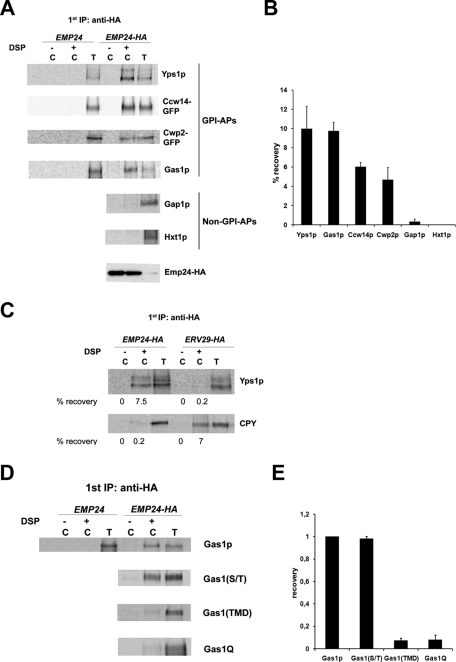
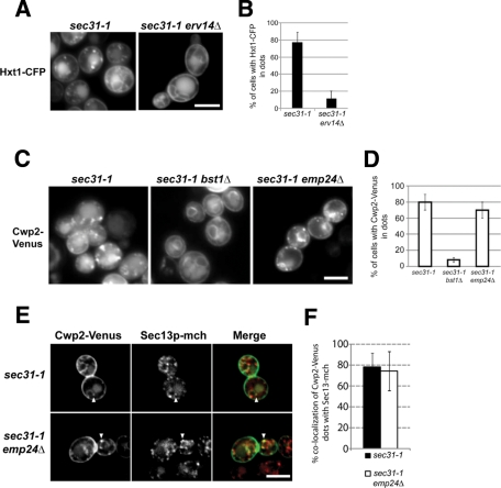
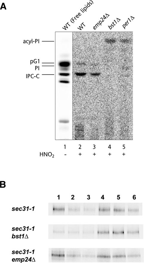
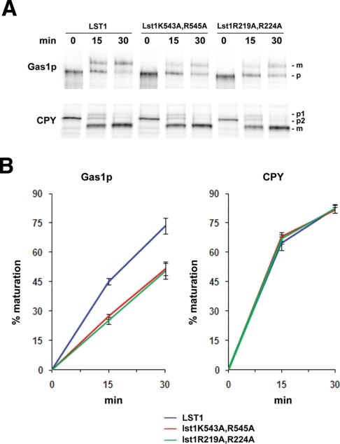
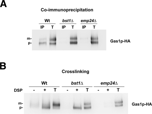
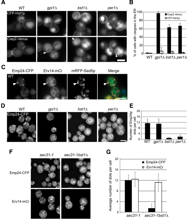
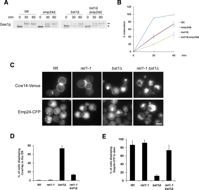
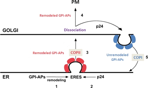
Glycosylphosphatidylinositol (GPI)-anchored proteins are secretory proteins that are attached to the cell surface of eukaryotic cells by a glycolipid moiety. Once GPI anchoring has occurred in the lumen of the endoplasmic reticulum (ER), the structure of the lipid part on the GPI anchor undergoes a remodeling process prior to ER exit. In this study, we provide evidence suggesting that the yeast p24 complex, through binding specifically to GPI-anchored proteins in an anchor-dependent manner, plays a dual role in their selective trafficking. First, the p24 complex promotes efficient ER exit of remodeled GPI-anchored proteins after concentration by connecting them with the COPII coat and thus facilitates their incorporation into vesicles. Second, it retrieves escaped, unremodeled GPI-anchored proteins from the Golgi to the ER in COPI vesicles. Therefore the p24 complex, by sensing the status of the GPI anchor, regulates GPI-anchored protein intracellular transport and coordinates this with correct anchor remodeling.


Figures

Figure 1:
Figure 1:
The p24 complex associates specifically with the GPI-anchored proteins through the GPI anchor. (A) Emp24p can be cross-linked specifically to GPI-anchored proteins in the ER. Pulse-radiolabeled yeast cells from EMP24-HA-tagged and untagged strains were converted to perforated spheroplasts and incubated with (+) and without (–) DSP. The samples were denatured and immunoprecipitated with anti-HA antibody and reprecipitated with antibody against the designated protein (labeled C). Five percent of the DSP-exposed spheroplasts were immunoprecipitated with antibodies against GPI-anchored proteins or non–GPI-anchored proteins to use as a standard (labeled T) for recovery. Samples were incubated with 5% β-mercaptoethanol, analyzed by SDS–PAGE, and visualized using a Phosphorimager. Emp24-HA was detected by immunoblot. (B) Quantification of several experiments described in A. The graph plots the average percentage of the recovery of different secretory proteins. GPI-APs, GPI-anchored proteins. (C) Erv29p can be cross-linked to CPY but not to GPI-anchored proteins. Spheroplasts obtained from Emp24-HA and Erv29-HA strains were incubated with (+) or without (–) DSP, denatured, immunoprecipitated with anti-HA antibody, and then reprecipitated with antibody against the Yps1p or CPY and processed as earlier. The percentage recovery of cross-linked Yps1p and CPY is shown. (D) Emp24p can be cross-linked to GPI-anchored proteins through the GPI anchor. Spheroplasts obtained from Emp24-HA cells expressing different constructs of the Gas1p were treated with (+) or without (–) DSP, denatured, immunoprecipitated with anti-HA antibody, and then reprecipitated with antibody against the Gas1p and processed as earlier. (E) Quantification of several experiments described in D. The graph plots the average percentage of the recovery of different Gas1p mutant constructs normalized relative to the recovery of wild-type Gas1p.
Figure 2:
Figure 2:
Emp24p is not required for GPI-anchored cargo sorting and concentration into ERES. (A) Fluorescence micrographs of live sec31-1 and sec31-1 erv14Δ cells expressing Hxt1-CFP at 37ºC. Raw images. (B) Quantification of several micrographs described in A. The graph plots the average percentage of the cells, for which Hxt1-CFP is found in dot-like structures. n, number of cells plotted; 74 ≤ n ≤ 89. (C) Fluorescence micrographs of live sec31-1, sec31-1 bst1Δ, and sec31-1 emp24Δ cells expressing Cwp2-Venus at 37ºC. Raw images. (D) Quantification of several micrographs described in C. The graph plots the average percentage of the cells, for which Cwp2-Venus is found in dot-like structures. n, number of cells plotted; 74 ≤ n ≤ 89. (E) Fluorescence micrographs of live sec31-1 and sec31-1 emp24Δ cells expressing Cwp2-Venus (green) and Sec13-mCh (red) at 37°C. White arrowheads, colocalizing dots. Deconvoluted images by 10 iterations. (F) Quantification of several micrographs described in E. The graph displays the means of the percentage of colocalization per cell of Cwp2-Venus dots with Sec13-mCh dots in sec31-1 (black bars, n = 36) and in sec31-1 emp24Δ (white bars, n = 55). Scale bar, A, C, E, 5 μm.
Figure 3:
Figure 3:
The p24 complex is not required for anchor remodeling or DRM partition of GPI-anchored proteins in the ER. (A) Lipid remodeling of the GPI anchor is normal in emp24Δ cells. Wild-type, emp24Δ, bst1Δ, and per1Δ strains were labeled with [3H]myo-inositol for 2 h at 25C. The labeled PI moieties were prepared from GPI-anchored proteins and analyzed by TLC using the solvent system 55:45:10 chloroform/methanol/0.25% KCl. Lipids extracted from wild-type cells (lane 1) were used as a standard. pG1, phosphatidylinositol with a C26:0 fatty acid in sn-2 position; PI, phosphatidylinositol; IPC-C, inositolphosphorylceramide consisting of 4-hydroxysphinganine and a hydroxylated C26:0 fatty acid (Fujita et al., 2006a); acyl-PI, inositol-acylated PI (Ghugtyal et al., 2007) (B) GPI-anchored proteins are associated with DRMs at the ER level in emp24Δ cells. DRM association of the Gas1p in the ER was analyzed using sec31-1, sec31-1 bst1Δ, and sec31-1 emp24Δ cells, which were previously pulse labeled and chased at 37ºC. The cells were broken with glass beads and subjected to TX-100 extraction and density gradient centrifugation. Six fractions were collected and analyzed by immunoprecipitation with antibodies against Gas1p.
Figure 4:
Figure 4:
The disruption of the p24 protein–binding site on the specialized COPII subunit Lst1p specifically impairs the efficient ER-to-Golgi transport of Gas1p. (A) Pulse-chase analysis of the ER-to-Golgi transport in the deletion strain lst1Δ expressing wild-type Lst1p or the mutant forms Lst1K543A,R545A and Lst1R219,224A. Proliferating cells were radiolabeled for 5 min, chased for the indicated times at 24°C, and lysed. Gas1p and CPY were immunoprecipitated, resolved by SDS–PAGE, and analyzed by Phosphorimager. Gas1p-p, ER-precursor form; -m, Golgi form. CPY-p2, ER-precursor form; -p1, Golgi precursor form; -m, mature form. (B) Quantification of several experiments described in A. The graph plots the average percentage of Gaslp and CPY maturation in lst1Δ strain expressing wild-type Lst1p or Lst1p mutants.
Figure 5:
Figure 5:
Emp24p can bind both remodeled and unremodeled GPI-anchored proteins. (A) Native coimmunoprecipitation assay between Emp24p and Gas1p. Enriched ER fractions (wild-type, bst1Δ, and emp24Δ mutant cells expressing Gas1-HA) were solubilized in 1% digitonin and analyzed by native immunoprecipitation (IP) with anti-Emp24p antibody followed by immunoblotting with anti-HA peroxidase antibody. Totals (T) represent a fraction of the solubilized input material. (B) Cross-linking assay between Emp24p and Gas1p. Spheroplasts from wild-type, bst1Δ, and emp24Δ mutant cells were incubated with (+) and without (−) DSP, denatured, and immunoprecipitated with anti-Emp24p antibody, followed by immunoblotting with anti-HA peroxidase antibody. Totals (T) represent a fraction of the solubilized input material. Gas1p: p, ER-precursor form; m, Golgi form.
Figure 6:
Figure 6:
Emp24p is relocalized from the Golgi to the ER in remodeling mutants. (A) Selective defect in the ER export of GPI-anchored proteins in remodeling mutants. Live images of wild-type, gpi1Δ, bst1Δ, and per1Δ expressing Hxt1-CFP and Cwp2-Venus at 30°C. (B) Quantification of several micrographs described in A. The graph plots the average percentage of cells displaying Cwp2-Venus (black bars) and Hxt1-CFP (white bars) in the ER. n, number of cells plotted; 37 ≤ n ≤ 53. (C) Live images of wild-type cells expressing Emp24-CFP, Erv14-mCi, and mRFP-Sed5 at 30°C. (D) Emp24p localization depends on remodeling of GPI-anchored proteins. Live images of wild-type, gpi1Δ, bst1Δ, and per1Δ cells expressing Emp24-CFP at 30°C. (E) Quantification of several micrographs described in D. The graph plots the average number of Emp24-CFP dots per cell seen in the different strains. n, number of cells plotted; 46 ≤ n ≤ 63. (F) Emp24p is not incorporated into ERES in remodeling mutants. Fluorescence micrographs of live sec31-1 and sec31-1 bst1Δ cells expressing Emp24-CFP and Erv14-mCi at 37ºC. (G) Quantification of several micrographs described in F. The graph plots the average percentage of the sec31-1 and sec31-1 bst1Δ cells for which Emp24-CFP and Erv14-mCi are found in dot-like structures. n, number of cells plotted. 74 ≤ n ≤ 89. A, C, D, Raw images. Scale bar, 5 μm.
Figure 7:
Figure 7:
Efficient ER retention of unremodeled GPI-anchored proteins requires their recycling from the Golgi to the ER by the p24 complex. (A) The emp24Δ mutation partially suppresses the GPI-anchored protein transport defect in remodeling mutants. Pulse-chase analysis to follow the transport from ER to Golgi of Gas1p in wild-type and deletion strains. Proliferating cells were radiolabeled for 5 min, chased for the indicated times at 24°C, and lysed. Gas1p was immunoprecipitated, resolved by SDS–PAGE, and analyzed by Phosphorimager. ER (p) and Golgi (m) Gas1p forms are indicated. (B) Quantification of several experiments described in A. The graph plots the average percentage of Gas1p maturation in wild-type and deletion strains. (C) An active retrograde transport is required for ER retention of GPI-anchored proteins and ER redistribution of Emp24p in remodeling mutants. Live images of wild-type, ret1-1, bst1Δ, and ret1-1 bst1Δ expressing Ccw14-Venus or Emp24-CFP at 24°C. Raw images. Scale bar, 5 μm. (D) Quantification of several micrographs described in C. The graph plots the average percentage of cells displaying Ccw14-Venus in the ER. n, number of cells plotted; n ≥ 100. (E) Quantification of several micrographs described in D. The graph plots the average number of Emp24-CFP dots per cell seen in the different strains. n, number of cells plotted; n ≥ 100.
Figure 8:
Figure 8:
Model of the specific roles of the p24 complex in the trafficking of GPI-anchored proteins along the early secretory pathway in yeast. The p24 complex promotes the efficient ER export of fully remodeled GPI-anchored proteins by linking the proteins to the COPII coat at their specific ERES and prevents the progression of incompletely remodeled GPI-anchored proteins along the secretory pathway by recycling them back from Golgi to the ER in COPI vesicles. 1) GPI anchored proteins are concentrated and sorted into their specific ERES upon anchor remodeling. 2) p24 complex is efficiently recruited to these ERES due to its binding to fully remodeled GPI-anchored proteins. 3) The p24 complex acts as an adaptor by linking COPII components to the GPI-anchored protein at ERES, which might facilitate vesicle biogenesis. 4) On arrival to the Golgi, GPI-anchored proteins dissociate from the p24 complex. On their release, correctly remodeled GPI-anchored proteins can progress through the secretory pathway to be delivered to the plasma membrane. 5) Escaped unremodeled GPI-anchored proteins are retrotransported from Golgi to the ER by the p24 complex. GPI-APs, GPI-anchored proteins.

References

    1. Aguilera-Romero A, Kaminska J, Spang A, Riezman H, Muniz M. The yeast p24 complex is required for the formation of COPI retrograde transport vesicles from the Golgi apparatus. J Cell Biol. 2008;180:713–720. - PMC - PubMed
    1. Bagnat M, Keranen S, Shevchenko A, Simons K. Lipid rafts function in biosynthetic delivery of proteins to the cell surface in yeast. Proc Natl Acad Sci USA. 2000;97:3254–3259. - PMC - PubMed
    1. Belden WJ, Barlowe C. Erv25p, a component of COPII-coated vesicles, forms a complex with Emp24p that is required for efficient endoplasmic reticulum to Golgi transport. J Biol Chem. 1996;271:26939–26946. - PubMed
    1. Belden WJ, Barlowe C. Distinct roles for the cytoplasmic tail sequences of Emp24p and Erv25p in transport between the endoplasmic reticulum and Golgi complex. J Biol Chem. 2001;276:43040–43048. - PubMed
    1. Bonnon C, Wendeler MW, Paccaud JP, Hauri HP. Selective export of human GPI-anchored proteins from the endoplasmic reticulum. J Cell Sci. 2010;123:1705–1715. - PubMed

Publication types

MeSH terms