. 2011 Sep;31(9):2114-24.
doi: 10.1161/ATVBAHA.111.230573. Epub 2011 Jun 16.

RAGE-dependent activation of the oncoprotein Pim1 plays a critical role in systemic vascular remodeling processes

Affiliations

RAGE-dependent activation of the oncoprotein Pim1 plays a critical role in systemic vascular remodeling processes

Jolyane Meloche et al. Arterioscler Thromb Vasc Biol. 2011 Sep.

Abstract

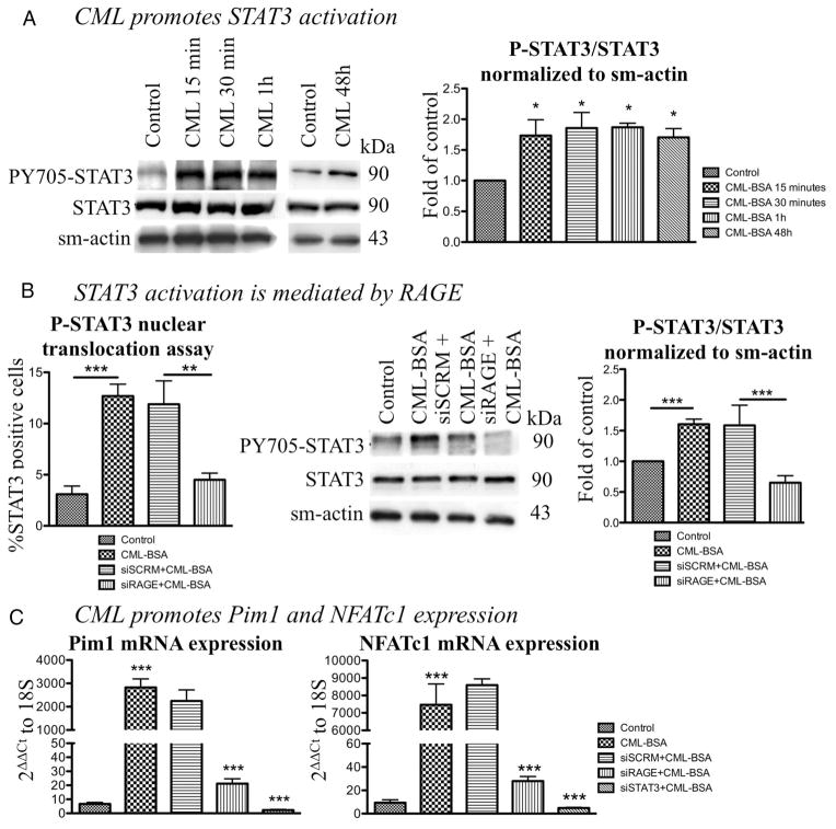
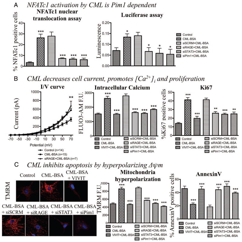
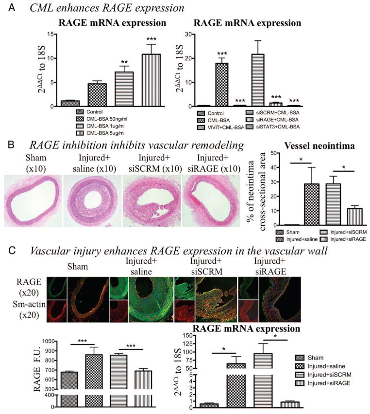
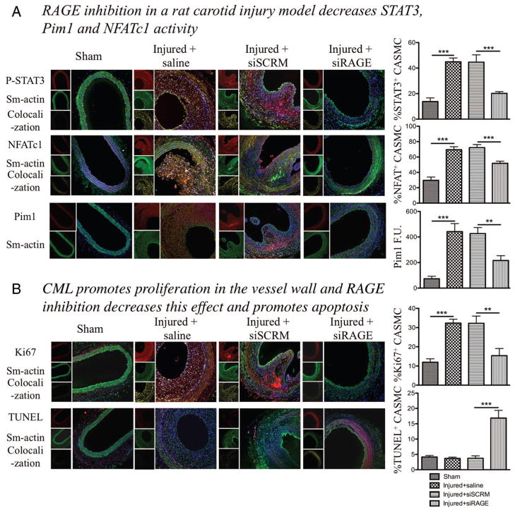
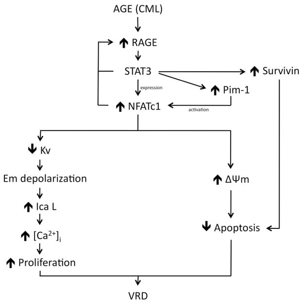
Objective: Vascular remodeling diseases (VRD) are mainly characterized by inflammation and a vascular smooth muscle cells (VSMCs) proproliferative and anti-apoptotic phenotype. Recently, the activation of the advanced glycation endproducts receptor (RAGE) has been shown to promote VSMC proliferation and resistance to apoptosis in VRD in a signal transducer and activator of transcription (STAT)3-dependant manner. Interestingly, we previously described in both cancer and VRD that the sustainability of this proproliferative and antiapoptotic phenotype requires activation of the transcription factor NFAT (nuclear factor of activated T-cells). In cancer, NFAT activation is dependent of the oncoprotein provirus integration site for Moloney murine leukemia virus (Pim1), which is regulated by STAT3 and activated in VRD. Therefore, we hypothesized that RAGE/STAT3 activation in VSMC activates Pim1, promoting NFAT and thus VSMC proliferation and resistance to apoptosis. Methods/Results- In vitro, freshly isolated human carotid VSMCs exposed to RAGE activator Nε-(carboxymethyl)lysine (CML) for 48 hours had (1) activated STAT3 (increased P-STAT3/STAT3 ratio and P-STAT3 nuclear translocation); (2) increased STAT3-dependent Pim1 expression resulting in NFATc1 activation; and (3) increased Pim1/NFAT-dependent VSMC proliferation (PCNA, Ki67) and resistance to mitochondrial-dependent apoptosis (TMRM, Annexin V, TUNEL). Similarly to RAGE inhibition (small interfering RNA [siRNA]), Pim1, STAT3 and NFATc1 inhibition (siRNA) reversed these abnormalities in human carotid VSMC. Moreover, carotid artery VSMCs isolated from Pim1 knockout mice were resistant to CML-induced VSMC proliferation and resistance to apoptosis. In vivo, RAGE inhibition decreases STAT3/Pim1/NFAT activation, reversing vascular remodeling in the rat carotid artery-injured model.

Conclusions: RAGE activation accounts for many features of VRD including VSMC proliferation and resistance to apoptosis by the activation of STAT3/Pim1/NFAT axis. Molecules aimed to inhibit RAGE could be of a great therapeutic interest for the treatment of VRD.


Figures

Figure 1
Figure 1
Nε-(carboxymethyl)lysine-bovine serum albumin (CML-BSA) enhances advanced glycation endproducts receptor (RAGE) expression and promotes STAT3/Pim1/NFATc1 axis. A, CML-BSA promotes STAT3 activation after only a 15-minutes exposition and its activation is maintained for 48 hours (1.5-fold activation; immunoblot, n=3, P<0.05). B, STAT3 activation is measured by nuclear translocation assay (n=50 CASMC/experiment for 4 experiments, P<0.01). CML-BSA induces a 80% increase in STAT3 activation and RAGE blockade (by siRNA 20 nmol/L) decreases the activation by 60%. We also confirmed that STAT3 activation was mediated by RAGE by immunoblot, showing that CML-BSA induces a 1.6-fold increase and RAGE blockade decreases STAT3 activation (n=4, P<0.01). C, CML-BSA triggers Pim1 and NFATc1 expression (800-fold increase; qRT-PCR, n=4, P<0.001). RAGE and STAT3 inhibition by siRNA induce a 150- and 300-fold decrease in Pim1 and NFATc1 expression, respectively (qRT-PCR).
Figure 2
Figure 2
Nε-(carboxymethyl)lysine-bovine serum albumin (CML-BSA) enhances Pim1 and nuclear factor of activated T-cells (NFAT)c1 expression and activation, which promotes cell proliferation and inhibits apoptosis in human carotid artery smooth muscle cells. A, CML-BSA also promotes NFATc1 activation, ie, translocation to the nucleus, through a RAGE/STAT3/Pim1-dependent mechanism (over 30% increase; immunofluorescence, n=50 CASMC/experiment for 4 experiments, P<0.001), also measured by luciferase assay (n=5, P<0.05). B, CML-BSA decreases K+ current (n=7 per group, P<0.01), which promotes intracellular calcium entry measured by FLUO3-AM by immunofluorescence (1.7-fold increase, n=50 CASMC/experiment for 5 experiments, P<0.001), which enhances cell proliferation. Cell proliferation was measured by activation of proliferation factor Ki67 (25% increase, n=50 CASMC/experiment for 5 experiments, P<0.001). To calculate the percentage of proliferation: cells positive for proliferation factors activated divided by total number of cell [visualized by 4,6-diamidino-2-phenylindole (DAPI)]. C, CML-BSA hyperpolarizes mitochondrial membrane potential measured by tetramethylrhodamine methyl ester (1.9-fold increase, n=50 CASMC/experiment for 5 experiments, P<0.001) which blocks cell death. CML-BSA inhibits apoptosis in CASMCs put in 0.1% of fetal bovine serum to stimulate apoptosis in normal cells (starvation) (n=50 CASMC/experiment for 5 experiments, P<0.05). Apoptosis was measured by calculating the percentage of positive AnnexinV cells, divided by total number of cell (visualized by DAPI). CML-BSA induced apoptosis resistance is mediated by RAGE/STAT3/Pim1/NFAT pathway, showed by an increase of apoptosis when either one of these effectors is blocked (by VIVIT 4 μmol/L or siRNA 20 nmol/L).
Figure 3
Figure 3
Enhanced advanced glycation endproducts receptor (RAGE) expression stimulates vascular remodeling. A, Nε-(carboxymethyl)lysine-bovine serum albumin (CML-BSA) enhances RAGE expression in a dose-dependent manner (measured by qRT-PCR normalized with 18S; n=4, P<0.01). Furthermore, RAGE expression in enhanced through a positive feedback loop including STAT3 and nuclear factor of activated T-cells (NFAT) because their blockade (siSTAT3 20 nmol/L and VIVIT 4 μmol/L, respectively) reduces RAGE expression (qRT-PCR, n=4, P<0.001). B, After H&E staining, neointima cross-sectional areas were measured. RAGE blockade showed decreased neointima area compared with injured carotids (55% decrease, n=5 per group, P<0.05). C, RAGE expression is enhanced in the injured vascular wall and siRAGE efficiency are shown by immunofuorescence, which corresponds to RAGE protein expression (n=5 per group, P<0.001) and qRT-PCR (n=5 per group, P<0.05).
Figure 4
Figure 4
Nε-(carboxymethyl)lysine-bovine serum albumin (CML-BSA) injected in rats stimulates the STAT3/Pim1/NFATc1 pathway and enhances vascular smooth muscle cells proliferation. A, CML-BSA promotes STAT3 activation through advanced glycation endproducts receptor (RAGE) in the vascular wall (immunofluorescence, n=5 rats per group, P<0.001). STAT3 activation correlates with nuclear factor of activated T-cells (NFAT) c1 activation (immunofluorescence, n=5 rats per group, P<0.001). Both STAT3 and NFAT are transcription factors and are present in the nucleus when activated. Thus, colocalization between either P-STAT3 or NFATc1 (red) and the nucleus blue, is showed by a yellow pattern, which is decreased when carotids were treated with RAGE siRNA. Pim1 expression is also enhanced in injured carotid and is inhibited when RAGE is blocked. Pim1 quantification was measured by fluorescence quantification (immunofluorescence, n=5 rats per group, P<0.01). B, RAGE/STAT3/Pim1/NFATc1 axis is responsible of cell proliferation, measured by Ki67 (immunofluorescence, n=5 rats per group, P<0.01). RAGE inhibition increases apoptosis in the vessel wall, measured by TUNEL (immunofluorescence, n=5 rats per group, P<0.01). For all staining, to calculate percentage of activation or proliferation, cells that had positive staining in nucleus were considered positive, which was divided by the total amount of cell (counted with DAPI). Again, yellow pattern shows colocalization between proliferation and apotosis factors (red) and the nucleus (blue).
Figure 5
Figure 5
Suggested pathway. Advanced glycation end product (AGE) molecules such as Nε-(carboxymethyl)lysine-bovine serum albumin (CML-BSA) activate STAT3 through their receptor advanced glycation endproducts receptor (RAGE). STAT3 is responsible for enhancing Survivin, Pim1, and nuclear factor of activated T-cells (NFAT) expressions. Pim1 is responsible of NFAT activation and NFAT stimulates proliferation through a decrease in Kv channel, which causes cell membrane (Em) depolarization. This depolarization causes a calcium intake through opening voltage-gated calcium channel known as long-lasting voltage-gated Ca channel ICaL which stimulates proliferation. On the other end, NFAT inhibits apoptosis by mitochondria membrane hyperpolarization. Survivin helps maintain resistance to apoptosis. Enhanced proliferation and resistance to apoptosis are responsible of vascular remodeling disease phenotype.

References

    1. Bonnet S, Paulin R, Sutendra G, Dromparis P, Roy M, Watson KO, Nagendran J, Haromy A, Dyck JR, Michelakis ED. Dehydroepiandrosterone reverses systemic vascular remodeling through the inhibition of the Akt/GSK3-{beta}/NFAT axis. Circulation. 2009;120:1231–1240. - PubMed
    1. Lambert CM, Roy M, Robitaille GA, Richard DE, Bonnet S. HIF-1 inhibition decreases systemic vascular remodelling diseases by promoting apoptosis through a hexokinase 2-dependent mechanism. Cardiovasc Res. 88:196–204. - PubMed
    1. Doran AC, Meller N, McNamara CA. Role of smooth muscle cells in the initiation and early progression of atherosclerosis. Arterioscler Thromb Vasc Biol. 2008;28:812– 819. - PMC - PubMed
    1. Libby P, Clinton SK. Cytokines as mediators of vascular pathology. Nouv Rev Fr Hematol. 1992;34 (Suppl):S47–S53. - PubMed
    1. Schwartz LB, Radic ZS, O’Donohoe MK, Mikat EM, McCann RL, Hagen PO. Saphenous vein endothelium-dependent relaxation in patients with peripheral vascular disease. Ann Vasc Surg. 1992;6:425– 432. - PubMed

Publication types