. 2011 Jun 17;12(8):804-10.
doi: 10.1038/embor.2011.102.

The role of trimerization in the osmoregulated betaine transporter BetP

Affiliations

The role of trimerization in the osmoregulated betaine transporter BetP

Camilo Perez et al. EMBO Rep. .

Abstract

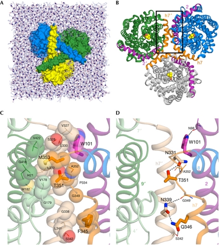
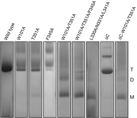
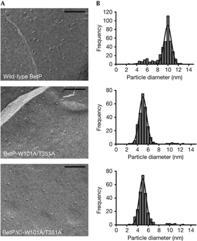
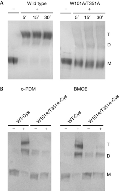
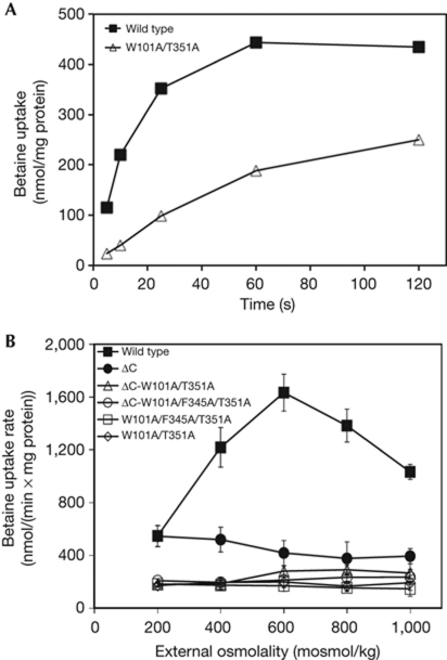
The osmoregulated betaine transporter BetP is a stable trimer. Structural studies have shown that individual protomers can adopt distinct transport conformations, implying a functional role for the trimeric state in transport, although the role of trimerization in regulation is not yet understood. We designed putative monomeric mutants by molecular-dynamics simulations and in silico alanine-scanning mutagenesis. Several mutants including BetP-W101A/T351A were monomeric in detergent as well as in the membrane, as shown by blue native gel electrophoresis, crosslinking and electron microscopy. This monomeric form retains the ability to accumulate betaine, but is no longer regulated by hyperosmotic shock.


Conflict of interest statement

The authors declare that they have no conflict of interest.

Figures

Figure 1
Figure 1
The structure of BetP and the simulation system. (A) The BetP trimer (surface representation) in a POPG lipid bilayer (sticks, with P atoms as spheres), viewed from the cytoplasm, showing the way in which the carboxy-terminal helices crossover to adjacent protomers. Water molecules and ions are omitted for clarity. (B) View of the BetP trimer from the periplasm. Helices at the trimer interface are shown in purple (TM2) or orange (h7); those belonging to the upper-left protomer are labelled 2′ and h7′, respectively. Bound betaine molecules are shown (spheres). (C,D) Close-up view of the protomer interface in a snapshot of a molecular-dynamics simulation, in the region marked with a box in (B), highlighting hydrophobic (C) or polar (D) interactions. Helices of two adjacent protomers (green and blue in B) are shown: h7 (beige), TM9 (dark green), TM4 and TM7 (pale green) from one protomer (labelled h7′, 4′, 7′ and 9′), and h7/L7-8 (orange), TM2 (purple) and TM8 (blue) from the second protomer. Side chains of hot-spot residues identified using in silico alanine scanning are shown (thick sticks, large labels), along with their primary interaction partners (thin sticks), and relevant inter-protomer hydrogen bonds are indicated (dashed lines). Residues involved in hydrophobic interactions are shown as spheres (C).
Figure 2
Figure 2
Mobility of wild-type BetP and mutants in BN–PAGE. Gel lanes are shown separately for viewing purpose; the complete gel and the molecular marker are shown in supplementary Fig S2 online. BN–PAGE, blue native polyacrylamide gel electrophoresis; D, dimer; M, monomer; T, trimer.
Figure 3
Figure 3
Results of freeze-fracture electron microscopy. (A) Electron micrographs of membrane-reconstituted wild-type BetP and mutants. Scale bar, 100 nm. (B) Distribution, that is, number of particles counted as a function of particle size. The line in each plot corresponds to a fit to a Gaussian function.
Figure 4
Figure 4
Crosslinking of BetP. (A) Glutaraldehyde crosslinking of detergent solubilized wild-type BetP and BetP-W101A/T351A, shown in a Coomassie-stained 10% SDS–PAGE. Samples were incubated with 0.13% glutaraldehyde (+) for the indicated times, up to 30 min. The control is indicated by −. (B) Western blot of BetP-D97C/S328C (WT-Cys) and BetP-D97C/W101A/S328C/T351A (W101A/T351A-Cys) in membrane vesicles of Escherichia coli cells. + indicates incubation with 5 mM of o-phenylenedimaleimide (o-PDM) or bis-maleimidoethane (BMOE). D, dimer; M, monomer; PAGE, polyacrylamide gel electrophoresis; T, trimer; WT, wild type.
Figure 5
Figure 5
Measurement of [14C]-betaine uptake rates in proteoliposomes. (A) Representative uptake of [14C]-betaine in proteoliposomes with wild-type BetP or the mutant BetP-W101A/T351A at 400 mosml/kg. (B) The wild-type BetP activation profile is compared with those of BetPΔC, BetP-W101A/T351A, BetP-W101A/F345A/T351A, BetPΔC-W101A/T351A and BetPΔC-W101A/F345A/T351A. Each value represents the average of three independent measurements. Error bars indicate the standard deviation.

References

    1. Eicher T, Brandstätter L, Pos KM (2009) Structural and functional aspects of the multidrug efflux pump AcrB. Biol Chem 390: 693–699 - PubMed
    1. Faraldo-Gómez JD, Smith GR, Sansom MS (2002) Setting up and optimization of membrane protein simulations. Eur Biophys J 31: 217–227 - PubMed
    1. Farwick M, Siewe RM, Kramer R (1995) Glycine betaine uptake after hyperosmotic shift in Corynebacterium glutamicum. J Bacteriol 177: 4690–4695 - PMC - PubMed
    1. Forrest L, Krämer R, Ziegler C (2011) The structural basis of secondary active transport mechanisms. Biochim Biophys Acta 1807: 167–188 - PubMed
    1. Herz K, Rimon A, Jeschke G, Padan E (2009) β-Sheet-dependent dimerization is essential for the stability of NhaA Na+/H+ antiporter. J Biol Chem 284: 6337–6347 - PubMed

Publication types

MeSH terms