. 2011 Jul;136(1):103-15.
doi: 10.1007/s00418-011-0826-y. Epub 2011 Jun 17.

Air-liquid interface cultures enhance the oxygen supply and trigger the structural and functional differentiation of intestinal porcine epithelial cells (IPEC)

Affiliations

Air-liquid interface cultures enhance the oxygen supply and trigger the structural and functional differentiation of intestinal porcine epithelial cells (IPEC)

Constanze Nossol et al. Histochem Cell Biol. 2011 Jul.

Abstract

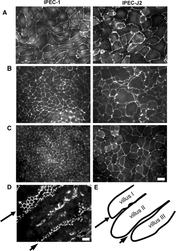
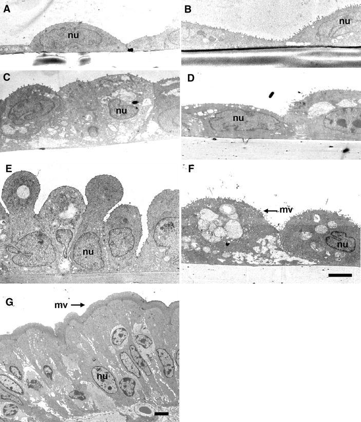
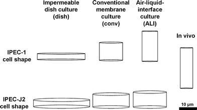
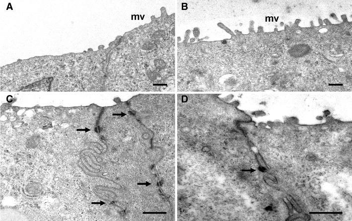
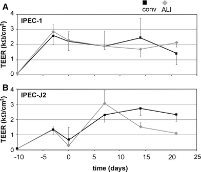
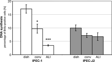
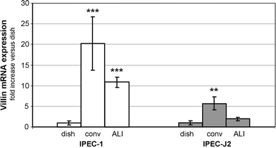
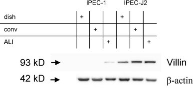
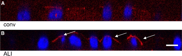
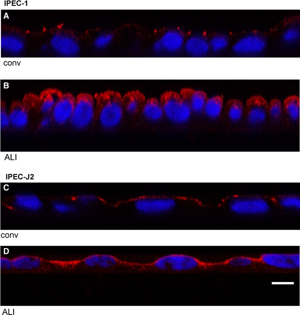
The specific function of the epithelium as critical barrier between the intestinal lumen and the organism's internal microenvironment is reflected by permanent maintenance of intercellular junctions and cellular polarity. The intestinal epithelial cells are responsible for absorption of nutritional components, facing mechanical stress and a changing oxygen supplementation via blood stream. Oxygen itself can regulate the barrier and the absorptive function of the epithelium. Therefore, we compared the dish cell culture, the transwell-like membrane culture and the oxygen enriched air-liquid interface (ALI) culture. We demonstrated strong influence of the different culture conditions on morphology and function of intestinal porcine epithelial cell lines in vitro. ALI culture resulted in a significant increase in cell number, epithelial cell layer thickness and expression as well as apical localisation of the microvilli-associated protein villin. Remarkable similarities regarding the morphological parameters were observed between ALI cultures and intestinal epithelial cells in vivo. Furthermore, the functional analysis of protein uptake and degradation by the epithelial cells demonstrated the necessity of sufficient oxygen supply as achieved in ALI cultures. Our study is the first report providing marked evidence that optimised oxygen supply using ALI cultures directly affects the morphological differentiation and functional properties of intestinal epithelial cells in vitro.


Figures

Fig. 1
Fig. 1
Influence of cell culture method on morphology and expression of tight junction protein ZO-1 in intestinal porcine epithelial cell lines IPEC-1 and IPEC-J2. Immunfluorescence staining of cells cultured on (a) impermeable dish (dish), (b) porous membrane (1-μm pore size) applying a conventional culture procedure (conv) and (c) porous membrane applying air–liquid-interface (ALI) culture conditions with enriched access to oxygen. d Section of porcine intestinal mucosa showing ZO-1 distribution in three villi in xy layer (flat section villus 1, arrow) and in zy layer (villus II and III, cross-section, arrowhead). e Low magnification scheme of section d. Scale bar 20 μm
Fig. 2
Fig. 2
Ultrastructural analysis of IPEC-1 and IPEC-J2 cell lines cultured with different supports and conditions. IPEC-1 cells (a, c, e) and IPEC-J2 cells (b, d, f) were cultured conventionally on impermeable support (a, b), on 1 μm membrane (c, d) and according to the ALI protocol on 1 μm membrane (e, f). A section of porcine jejunum is shown for comparison (g). Apical side of the cell layer is on the top. Microvilli structures (mv) are located on apical membrane (eg) and basolateral intercellular spaces are shown (cg). Nucleus (nu). Scale bar 4 μm (af)
Fig. 3
Fig. 3
Visualization of geometric parameters of IPEC-1 and IPEC-J2 cells as a function of cell culture condition. IPEC-1 and IPEC-J2 cells were cultured under different cell culture conditions as indicated. Average cell diameter and cell thickness were used to calculate a hypothetical cell volume representing characteristic cell shape under the given culture condition. Scale bar 10 μm
Fig. 4
Fig. 4
Ultrastructural view of apical side of membrane cultured IPEC-1 and IPEC-J2 cells. Electron microscopical analysis of apical side of cultured intestinal cell lines exhibit low degree of microvilli (mv) in IPEC-1 cells (a) in comparison to IPEC-J2 cells (b). Characteristic tight junction structures (arrows) between adjacent cells were found under all culture conditions in IPEC-1 (c) and IPEC-J2 (d) cells. Scale bar 0.5 μm
Fig. 5
Fig. 5
TEER measurement of IPEC-1 and IPEC-J2 cells cultured on conventional membrane and as ALI system. IPEC-1 (a) and IPEC-J2 (b) cells were seeded (day −10) on cell culture inserts with 1-μm pore size (12-well format) and TEER was regularly monitored (TEER kΩ/cm2 and SEM.). At day 0 cultures were switched to ALI culture mode (ALI) or were further handled conventionally (conv). IPEC-1 (a) cells formed cell layers with TEER > 1 kΩ/cm2 at day 0 and maintained the electrical dense structure throughout the cultivation. IPEC-J2 (b) cell layers were around 1 kΩ/cm2 at day 0, but exceeded this value during the following cultivation
Fig. 6
Fig. 6
DNA synthesis of IPEC-1 and IPEC-J2 cells cultured on dish, on conventional membrane and as ALI system. Proliferation of IPEC-1 and IPEC-J2 cells was quantified by detection of BrdU incorporation on single cell level. Percentage of BrdU-positive cell nuclei were estimated on dish, membrane (conv) and ALI cultures. Percentage was calculated from at least three independent experiments ± S.E.M. Significant changes are indicated as *p ≤ 0.01 and ***p ≤ 0.001 versus cells cultured on dish
Fig. 7
Fig. 7
Comparison of villin mRNA of IPEC-1 and IPEC-J2 cells cultured on dish, on conventional membrane or as ALI system. IPEC-1 and IPEC-J2 cells were cultured for 21 days on impermeable surface (dish) and on 1 μm membrane in conventional (conv) or ALI (ALI) mode. Levels of mRNA of villin were detected by qRT-PCR and related to mRNA levels detected in corresponding dish culture. Membrane and ALI cultures significantly increased villin mRNA level in IPEC-1. In IPEC-J2 conventional membrane culture significantly increased villin mRNA level, but ALI mode was without significant effect. Increase of mRNA level of villin was calculated from at least six independent experiments ± S.E.M. Significant changes in ratio in comparison to lowest mRNA level are indicated as **p ≤ 0.01 and ***p ≤ 0.001 versus cells cultured on dish
Fig. 8
Fig. 8
Comparison of villin protein expression in IPEC-1 and IPEC-J2 cells cultured on dish, on conventional membrane or as ALI system. IPEC-1 and IPEC-J2 cells were cultured for 21 days on impermeable surface (dish) and on 1 μm membrane in conventional or ALI mode. Levels of villin protein were detected by western blotting using mouse monoclonal anti-villin antibody and chemoluminescence detection. GAPDH was used as a loading control. In IPEC-1 villin was detectable in membrane cultures of the ALI mode only. IPEC-J2 expressed villin protein in all culture modes with slight enhanced protein levels in membrane and ALI cultures
Fig. 9
Fig. 9
Comparison of villin protein expression in IPEC-J2 cells cultured on conventional membrane or as ALI system. IPEC-J2 cells were cultured on 1 μm membranes in conventional (a) and ALI culture mode (b). Villin was detected by immunfluorescence staining and distribution was visualized by confocal microscopy. Figures represent optical sections obtained after three-dimensional reconstructions. In the conventional culture mode, villin is detectable but homogenously distributed in the cell (a). In the ALI culture mode villin is localised at the apical pole of the plasma membrane (b arrows). A representative view is shown. Scale bar 5 μm
Fig. 10
Fig. 10
Uptake of fluorescence-labelled BSA structures in IPEC-1 and IPEC-J2 cell cultured on conventional membrane or as ALI system. IPEC-1 (a, b) and IPEC-J2 (c, d) cells were cultured on 1 μm membranes in conventional (a, c) and ALI culture mode (b, d). DyLight-labelled protein (BSA) was applied apically and confocal microscopy of fixed cells was done after 24 h. Figures represent orthogonal views obtained after three-dimensional reconstructions. The membrane is localised basal of the nuclei. Fluorescence signal was found on the apical border of conventional cultures (a, c) only. ALI cultured cells exhibit intracellular fluorescence (b, d). Scale bar 5 μm

References

    1. Bacallao R, Garfinkel A, Monke S, Zampighi G, Mandel LJ. ATP depletion: a novel method to study junctional properties in epithelial tissues. I. Rearrangement of the actin cytoskeleton. J Cell Sci. 1994;107(Pt 12):3301–3313. - PubMed
    1. Bader A, Hansen T, Kirchner G, Allmeling C, Haverich A, Borlak JT. Primary porcine enterocyte and hepatocyte cultures to study drug oxidation reactions. Br J Pharmacol. 2000;129:331–342. doi: 10.1038/sj.bjp.0703062. - DOI - PMC - PubMed
    1. Barnard JA, Warwick G. Butyrate rapidly induces growth inhibition, differentiation in HT-29 cells. Cell Growth Differ. 1993;4:495–501. - PubMed
    1. Basson MD, Turowski G, Emenaker NJ. Regulation of human (Caco-2) intestinal epithelial cell differentiation by extracellular matrix proteins. Exp Cell Res. 1996;225:301–305. doi: 10.1006/excr.1996.0180. - DOI - PubMed
    1. Brandtzaeg P, Tolo K. Mucosal penetrability enhanced by serum-derived antibodies. Nature. 1977;266:262–263. doi: 10.1038/266262a0. - DOI - PubMed

Publication types

LinkOut - more resources