

Review
doi: 10.1007/s12105-011-0274-y.
Epub 2011 Jun 18.
Oral elastofibromatous lesions: a review and case series
Affiliations
- PMID: 21681661
- PMCID: PMC3173539
- DOI: 10.1007/s12105-011-0274-y
Item in Clipboard
Review
Oral elastofibromatous lesions: a review and case series
Head Neck Pathol.
2011 Sep.
Abstract
Elastofibromas of the oral cavity are rare, with only 5 cases reported. In this paper, we present a series of five new cases of oral elastofibromatous lesions, occurring in 4 males and 1 female, with ages ranging from 33 to 76 years. The clinical differential diagnosis includes fibroepithelial polyp or fibroma, among other connective tissue tumours. Elastofibromas probably develop as reactive lesions, for which surgical treatment is definitive.
Figures
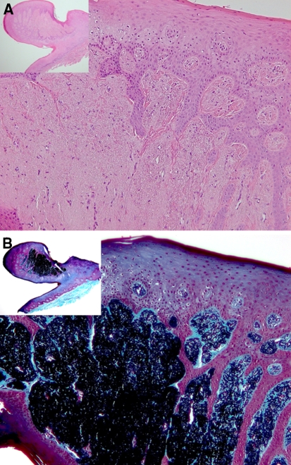
a Case 1: elastofibroma showing a raised nodular growth. (H&E stain; original magnification ×100; inset ×25). b Case 1: elastic fiber proliferation shown with MET stain (original magnification ×100; inset ×25)
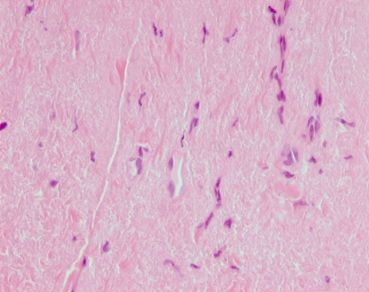
Case 2: proliferation of elastic fibers, resembling elastosis in many areas (H&E stain; original magnification ×400)
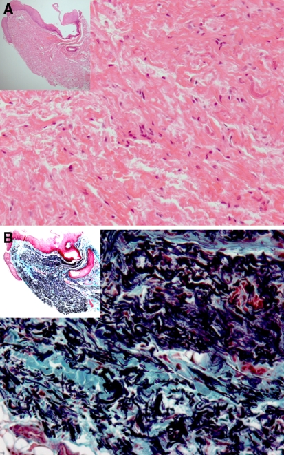
a Case 3: This elastofibroma was described clinically as a “leukoplakia”, and shows an apparent raised area microscopically (H&E stain;. original magnification ×100; inset ×25). b Case 3: Elastic fiber proliferation shown with MET stain (original magnification ×100; inset ×25)
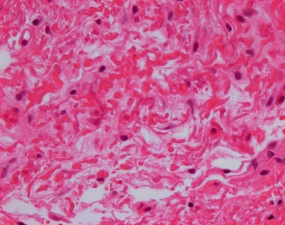
Case 3: Globular and occasional “serrated edge” configuration of elastic fibers (H&E stain; original magnification ×400)
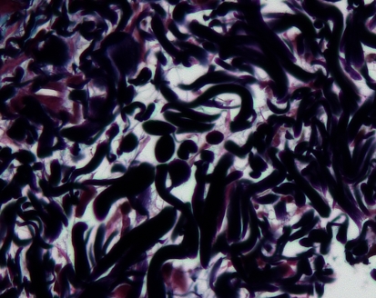
Case 4: Elastic fibers dominated, showing a mostly globular arrangement (MET stain; original magnification ×400)
Comment in
-
Elastofibroma oralis.Head Neck Pathol. 2011 Sep;5(3):259-60. doi: 10.1007/s12105-011-0299-2. Epub 2011 Sep 6. Head Neck Pathol. 2011. PMID: 21894499 Free PMC article. No abstract available.
References
Publication types
MeSH terms
LinkOut - more resources
Full Text Sources
Medical
