. 2011 Oct 15;519(15):2907-21.
doi: 10.1002/cne.22700.

Delayed synapse elimination in mouse levator palpebrae superioris muscle

Affiliations

Delayed synapse elimination in mouse levator palpebrae superioris muscle

Michael A Fox et al. J Comp Neurol. .

Abstract

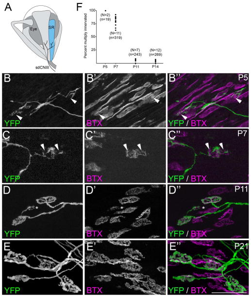
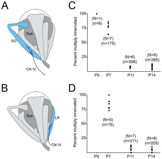
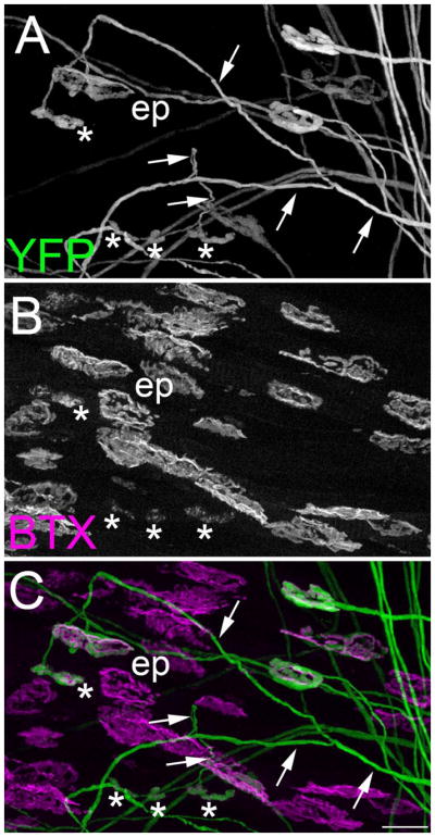
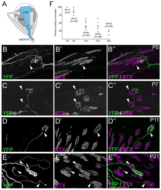
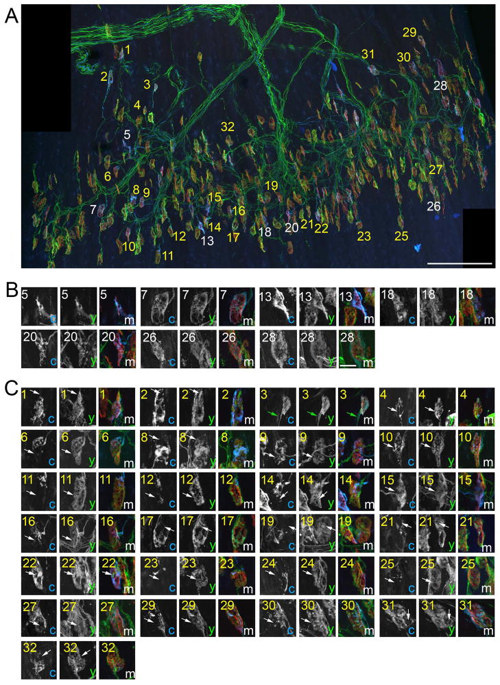
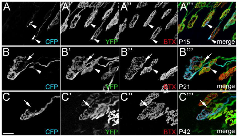
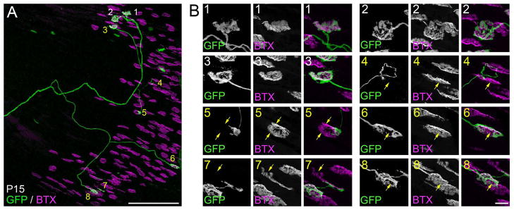
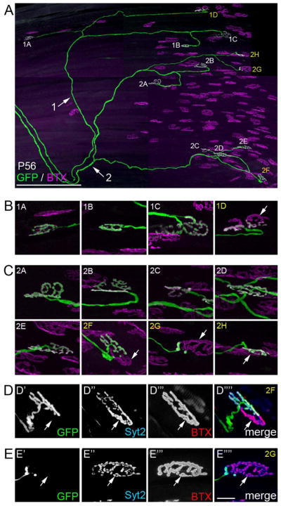
At birth, synaptic sites in developing rodent muscles are innervated by numerous motor axons. During subsequent weeks, this multiple innervation disappears as one terminal strengthens, and all the others are eliminated. Experimental perturbations that alter neuromuscular activity affect the rate of synaptic refinement, with more activity accelerating the time to single innervation and neuromuscular blockade retarding it. However, it remains unclear whether patterns of muscle use (driven by endogenous neuronal activity) contribute to the rate of synapse elimination. For this reason we examined the timing of supernumerary nerve terminal elimination at synapses in extraocular muscles (EOMs), a specialized set of muscles controlling eye movements. On the basis of their exceptionally high patterns of activity, we hypothesized that synaptic refinement would be greatly accelerated at these synapses. We found, however, that rates of synaptic refinement were only modestly accelerated in rectus and oblique EOMs compared with synapses in somite-derived skeletal muscle. In contrast to these results, we observed a dramatic delay in the elimination of supernumerary nerve terminals from synapses in the levator palpebrae superioris (LPS) muscle, a specialized EOM that initiates and maintains eyelid elevation. In mice, natural eye opening occurs at the end of the second postnatal week of development. Thus, although synapse elimination is occurring in most EOMs and somite-derived skeletal muscles, it appears to be dramatically delayed in a set of specialized eyelid muscles that remain immobile during early postnatal development.


Figures

Fig. 1
Fig. 1. Development of motor nerve terminals and AChR clusters at ‘en plaque’-type synapses in SR muscle
A. Schematic representation of SR muscle (in blue) and its innervation by the superior division of the oculomotor nerve (sdCNIII). B–E. YFP-expressing motor nerve terminals (green) were imaged in P5 (B), P7 (C), P11 (D) and P21 (E) SR muscles isolated from thy1:yfp line H mice. AChR clusters were simultaneously visualized by labeling with fluorescently-conjugated bungarotoxin (BTX; magenta). At early ages (B,C) YFP-containing motor nerve terminals failed to innervate the entire postsynaptic membrane, suggesting that synaptic sites remained multiply innervated at these ages. Arrowheads highlight AChR-rich regions of the postsynaptic membrane that were unoccupied by YFP-labeled nerve terminals. By P11 (D), few synaptic sites contacted by YFP-labeled axons appeared multiply innervated. Asterisk in D, highlights an ‘en grappe’-type AChR cluster contacted by the same motor axon that contacted 2 ‘en plaque’-type NMJs in this field of view. Scale bar = 50 μm. F. Numbers of multiply innervated NMJs were quantified at P5, P7, P11, and P14 from thy1:yfp line H SR muscles. Each black dot represents the percentage of YFP-labeled NMJs that were multiply innervated from a single muscle. N = number of muscles analyzed per age. n = total of number of NMJs analyzed per age.
Fig 2
Fig 2. Percent of multiply innervated ‘en plaque’-type NMJs in developing SO and LR muscles
A,B. Schematic representation of SO (A) and LR (B) muscles (in blue) and their innervation by CN IV and CN VI, respectively. Numbers of multiply innervated NMJs at P5, P7, P11, and P14 were quantified from SO (C), and LR (D) muscles isolated from thy1:yfp line H mice. Each black dot represents the percentage of YFP-labeled NMJs that were multiply innervated from a single muscle. N = number of muscles analyzed per age. n = total of number of NMJs analyzed per age.
Fig 3
Fig 3. “En plaque’- and ‘en grappe’-type AChR clusters were contacted by the same motor axons
YFP-expressing motor nerve terminals (A)(green) were imaged in P21 SR muscles isolated from thy1:yfp line H mice. AChR clusters were labeled with fluorescently-conjugated bungarotoxin (B)(BTX; magenta). A merged image is shown in C. Note that the YFP-labeled axon highlighted with arrows contacts both ‘en plaque’- (ep) and ‘en grappe’-type (asterisks) synaptic sites. Scale bars = 25 μm.
Fig 4
Fig 4. Development of nerve terminals and AChR clusters in LPS muscle
A. Schematic representation of LPS muscle (in blue) and its innervation by the superior division of the oculomotor nerve (sdCNIII). B–E. YFP-expressing motor nerve terminals (green) were imaged in P5 (B), P7 (C), P11 (D) and P21 (E) LPS muscles isolated from thy1:yfp line H mice. AChR clusters were simultaneously visualized by labeling with fluorescently conjugated bungarotoxin (BTX; magenta). At all ages, YFP-containing motor nerve terminals were observed that failed to innervate entire postsynaptic sites, indicating these synapses were multiply innervated. Arrowheads highlight AChR-rich regions of the postsynaptic membrane unoccupied by YFP-labeled nerve terminals. Scale bar = 50μm. F. Numbers of NMJs multiple innervated synapses in P5, P7, P11, P14 and P21 LPS muscles isolated from thy1:yfp line H mice. Each black dot represents the percentage of YFP-labeled NMJs that were multiply innervated from a single LPS muscle. N = number of LPS muscles analyzed per age. n = total of number of NMJs analyzed per age.
Fig 5
Fig 5. Multiple nerve terminals were observed at most synaptic sites in P10 LPS muscles
To conclusively determine synaptic sites in P10 LPS muscle were contacted by multiple axons, NMJs were imaged in thy1:yfp line 16; thy1:cfp line23 mice in which all motor axons were labeled with YFP and only a few were also labeled with CFP. A. A montage of high-resolution images encompassing the entire central end-plate band. AChR clusters were labeled with BTX (red). 32 NMJs contacted by a CFP-labeled axon were labeled with numbers: white numbers depict singly innervated NMJs; yellow numbers depict synapses contacted by both CFP-labeled axons and YFP-labeled (CFP-non-labeled) axons. B. High magnification images of singly innervated NMJs from (A). C. High magnification images of NMJs that are contacted by both a CFP-expressing axon and supernumerary YFP-expressing axons. Arrows in C highlight domains of these NMJs that are innervated by YFP-labeled (CFP-non-labeled) nerve terminals. In some cases although CFP-labeled terminals appeared to fully occupy a postsynaptic site, YFP-labeled (CFP-non-labeled) axons were still observed co-innervating these site (see green arrows in image number 3 of C). c = CFP; y = YFP; m = merge. Scale bar in A = 200 μm, in B = 10 μm for B,C.
Fig 6
Fig 6. Multiple nerve terminals were observed at synaptic sites in P15, P21 and P42 LPS muscles
NMJs were imaged from P15 (A), P21 (B) and P42 (C) LPS muscles from thy1:yfp line 16; thy1:cfp line23 transgenic mice in which all motor axons were labeled with YFP and only a few were also labeled with CFP. Note the segregation of axonal terminals into distinct domains of the postsynaptic membrane in P21 and P42 LPS muscles (B,C). Arrows highlight AChR-rich regions unoccupied by CFP-labeled motor nerve terminals. Arrowhead in B highlights a small portion of this synaptic site contacted by a CFP-labeled axon. Scale bar = 10 μm.
Fig 7
Fig 7. Reconstruction of a single LPS motor unit revealed a high degree of multiple innervation at P15
A. Confocal reconstruction of a single motor axon in a P15 thy1:gfp line S LPS muscle. Synaptic sites contacted by this axon are labeled with numbers. White numbers depict singly innervated synapses. Yellow numbers depict synapses in which GFP-labeled axons do not fully occupy all of the AChR-rich postsynaptic sites, indicating they are multiple innervated. B. High magnification images of synaptic sites contacted by the labeled motor axon shown in A. Yellow arrows highlight regions of AChR-rich postsynaptic membranes not contacted by the GFP-labeled axon. Scale bar in A = 250 μm and in B = 10 μm.
Fig 8
Fig 8. Reconstruction of two LPS motor units revealed multiply innervated synaptic sites persisted at P56
A. Confocal reconstruction of two motor axons innervating the same P56 LPS muscle from a thy1:gfp line S mouse. Each axon is labeled (with arrows and either a 1 or 2) as they course toward the end-plate band. Synaptic sites contacted by each axon are labeled with numbers that correspond to the axon innervating them and a letter. Sites labeled in white appear singly innervated; sites labeled in yellow synapses appear multiply innervated. B. High magnification images of synaptic sites in motor unit 1. Arrow indicates AChR-rich regions of a synaptic site not fully innervated by the GFP-labeled axon. C. High magnification images of synaptic sites in motor unit 2. Arrows indicates AChR-rich regions of synaptic sites not fully innervated by the GFP-labeled axon. D,E. After imaging, the LPS muscle was immunostained for synaptotagmin 2 (Syt2), a component of the presynaptic machinery within all motor nerve terminals. Immuno-labeling for Syt2 (blue) revealed that portions of the synaptic sites not occupied by the GFP-labeled were occupied by other axons (see arrows). Scale bar in A = 250 μm and in E = 10 μm for B–E.

Similar articles

Cited by

References

    1. Bach-y-rita P, Lennerstrand G. Absence of polyneuronal innervation in cat extraocular muscles. J Physiol. 1975;244(3):613–624. - PMC - PubMed
    1. Balice-Gordon RJ, Lichtman JW. In vivo observations of pre- and postsynaptic changes during the transition from multiple to single innervation at developing neuromuscular junctions. J Neurosci. 1993;13(2):834–855. - PMC - PubMed
    1. Balice-Gordon RJ, Chua CK, Nelson CC, Lichtman JW. Gradual loss of synaptic cartels precedes axon withdrawal at developing neuromuscular junctions. Neuron. 1993;11(5):801–815. - PubMed
    1. Balice-Gordon RJ, Lichtman JW. Long-term synapse loss induced by focal blockade of postsynaptic receptors. Nature. 1994;372(6506):519–524. - PubMed
    1. Benoit P, Changeux JP. Consequences of tenotomy on the evolution of multiinnervation in developing rat soleus muscle. Brain Res. 1975;99(2):354–358. - PubMed

Publication types

MeSH terms

Substances