. 2012 Feb;72(3):291-300.
doi: 10.1002/pros.21431. Epub 2011 Jun 16.

Aberrant expression of katanin p60 in prostate cancer bone metastasis

Affiliations

Aberrant expression of katanin p60 in prostate cancer bone metastasis

Xiangcang Ye et al. Prostate. 2012 Feb.

Abstract

Background: Katanin p60 is a microtubule-severing protein and is involved in microtubule cytoskeleton organization in both mitotic and non-mitotic processes. Its role in cancer metastasis is unknown.

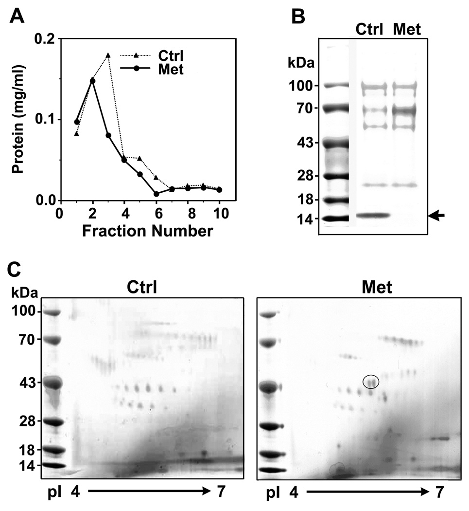
Methods: Differential protein profiles of bone marrow aspirates were analyzed by chromatography, electrophoresis, and mass spectrometry. Expression of katanin p60 in primary and metastatic prostate cancer was examined by immunohistochemistry. Cellular function of katanin p60 was further examined in prostate cell lines.

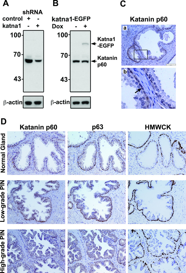
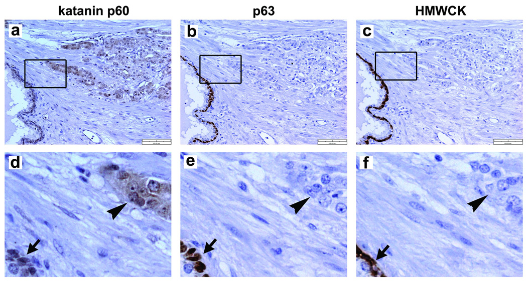
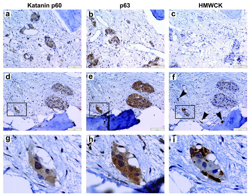
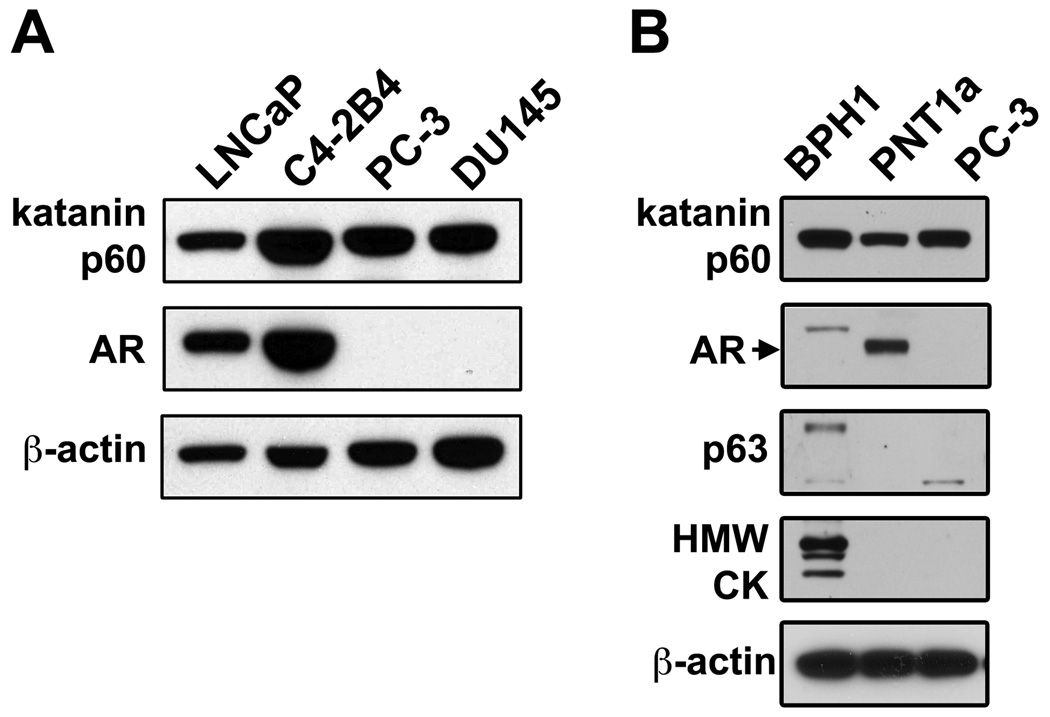
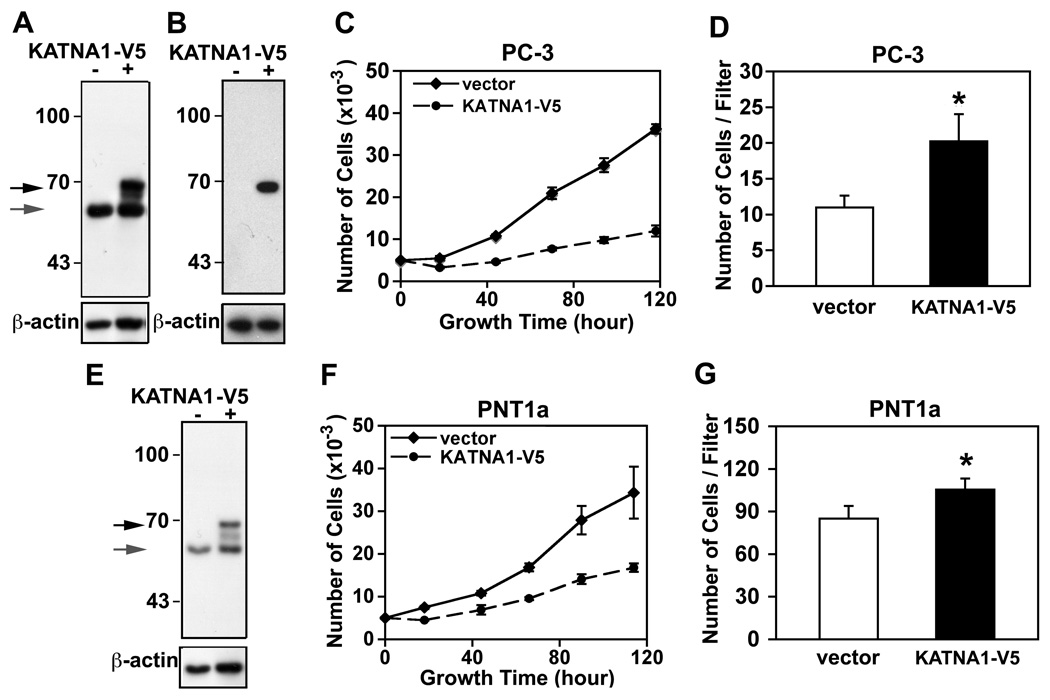
Results: In a proteomic profiling of bone marrow aspirates from men with prostate cancer, we found that katanin p60 was one of the proteins differentially expressed in bone metastasis samples. Immunohistochemical staining showed that katanin p60 was expressed in the basal cells in normal human prostate glands. In prostatic adenocarcinomas, in which the basal cells were absent, katanin p60 was expressed in the prostate cancer cells. In the specimens from bone metastasis, katanin p60 was detectable in the metastatic cancer cells. Strikingly, some of the metastatic cancer cells also co-expressed basal cell biomarkers including the tumor suppressor p53-homologous protein p63 and the high molecular weight cytokeratins, suggesting that the metastatic prostate cancer cells may have a basal cell-like phenotype. Moreover, overexpression of katanin p60 inhibited prostate cancer cell proliferation but enhanced cell migration activity.

Conclusions: Katanin p60 was aberrantly expressed during prostate cancer progression. Its expression in the metastatic cells in bone was associated with the re-emergence of a basal cell-like phenotype. The elevated katanin p60 expression may contribute to cancer cell metastasis via a stimulatory effect on cell motility.


Conflict of interest statement

CONFLICT OF INTEREST

The authors declared no potential conflicts of interest with respect to the authorship and/or publication of this article.

Figures

Figure 1
Figure 1
Identification of differentially expressed proteins in the bone marrow samples from men with or without bone metastasis from prostate cancer. A. Elution profiles of the pooled bone marrow supernatants with WGA-agarose affinity chromatography. Bone marrow supernatants from prostate cancer patients who did not have clinical evidence of bone metastasis served as control (Ctrl) for those with bone metastasis (Met). B. SDS-PAGE of the WGA-affinity purified fractions. Arrow indicates a decrease in the level of a 16 kDa protein in the Met samples. Mass spectrometry analysis identified it as hemoglobin β-chain. C. Two-dimensional gel electrophoresis of the proteins eluted from WGA-affinity chromatography. Two discernible protein spots in the Met samples are circled. One of them was identified to be katanin p60 by mass spectrometry analysis.
Figure 2
Figure 2
Validation of katanin p60 antibody and immunohistochemical staining of normal prostatic glands and prostatic intraepithelial neoplasia. A. Western blot with katanin p60 antibody showed a 60 kDa band that was reduced after knockdown by KATNA1-shRNA but not by control shRNA in 293T cells. B. Katanin p60 antibody recognized the 85 kDa katanin p60-EGFP fusion protein. Expression of katanin p60-EGFP was achieved by transfection with a pTRE-KATNA1-EGFP in a Tet-On 293T cell line and induction with 1 µg/ml Doxycycline (Dox) for 24 hours. C. Immunostaining of normal prostate tissues with katanin p60 antibody showed that katanin p60 protein was weakly detectable in the epithelial cells. A boxed region is enlarged in panel (b). Note that arrow points to katanin p60 staining in the basal cell layer. D. Immunostaining of katanin p60 and basal cell markers (i.e. p63 and HMWCK) in normal prostatic glands, low-grade and high-grade PIN.
Figure 3
Figure 3
Immunostaining of katanin p60 and basal cell biomarkers in prostatic carcinomas. (a) Elevated expression of katanin p60 was detectable in carcinomas. A representative sample (Greason grade 4) is shown. An enlarged area is shown in panel (d). Arrowhead indicates katanin p60 staining in the carcinoma cells. Arrow indicates katanin p60 staining in the basal cells in a neighboring normal gland. (b) An adjacent tissue section was stained with anti-p63 antibody clone 4A4. An enlarged area (e) showed the carcinoma was negative for p63 staining (arrowhead). (c) An additional adjacent tissue section was stained with anti-HMWCK antibody 34βE12. The enlarged image (f) showed the carcinoma was also negative of HMWCK staining (arrowhead), while the basal cells in the neighboring gland were positive for HMWCK staining (arrow).
Figure 4
Figure 4
Immunostaining of katanin p60 and the basal cell biomarkers in the bone metastasis of prostate cancer. (a and d) Expression of katanin p60 was detectable in the scattering metastatic cells and tumor foci in bone marrow cavities. Two independent samples were shown. The boxed area from (d) is enlarged in panel (g), in which katanin p60 was localized primarily in the cytoplasm. (b and e) In the adjacent tissue sections, the basal cell marker p63 was detected in the cytoplasm of metastatic cells and tumor foci. An enlarged area from (e) is shown in panel (h). (c and f) High molecule weight cytokeratins (HMWCK) were detected in a minor population of metastatic cells and small tumor foci in the additional adjacent tissue sections. Note the arrowheads point to scattered single prostate cancer cells (f) and the basal-like cells at the edge of a small tumor focus (i).
Figure 5
Figure 5
Expression of katanin p60, androgen receptor and basal cell markers in human prostate cell lines. A. Katanin p60 was detected by Western blotting in human prostate cancer cell lines. Androgen receptor was detected by Upstate PG-21 antibody. B. Expression of prostatic basal cell markers p63 and HMWCKs in BPH1, PNT1a and PC-3 cells were examined with p63-4A4 and CK-34βE12 antibodies, respectively.
Figure 6
Figure 6
Effect of katanin p60 overexpression on PC-3 and PNT1a cell growth and migration. A. Overexpression of V5-tagged katanin p60 in PC-3 cells by transient transfection of a pcDNA3.1D-KATNA1-V5-6xHis (KATNA1-V5). Recombinant protein and endogenous katanin p60 were detected by immunoblotting with anti-katanin p60 antibody. B. The recombinant protein was confirmed by anti-V5 epitope antibody. C. Overexpression of katanin p60 inhibits cell proliferation in PC-3 cells. Growth curves were recorded from the PC-3 cells transfected with an empty vector or a KATNA1-V5 expression construct. D. Cell migration properties of the transfected PC-3 cells were determined in transwell assays. Three independent experiments for each cell line were performed and a representative experiment with PC-3 cells is shown. *, P < 0.05. E. Recombinant and endogenous katanin p60 in PNT1a cells were detected by immunoblotting with anti-katanin p60 antibody. F. Overexpression of katanin p60 inhibited cell proliferation in PNT1a cells. G. Elevated katanin p60 levels induced an increase in cell migration in PNT1a cells. *, P < 0.05.

Similar articles

Cited by

References

    1. Bubendorf L, Schopfer A, Wagner U, Sauter G, Moch H, Willi N, Gasser TC, Mihatsch MJ. Metastatic patterns of prostate cancer: an autopsy study of 1,589 patients. Hum Pathol. 2000;31(5):578–583. - PubMed
    1. Shah RB, Mehra R, Chinnaiyan AM, Shen R, Ghosh D, Zhou M, Macvicar GR, Varambally S, Harwood J, Bismar TA, Kim R, Rubin MA, Pienta KJ. Androgen-independent prostate cancer is a heterogeneous group of diseases: lessons from a rapid autopsy program. Cancer Res. 2004;64(24):9209–9216. - PubMed
    1. McNally FJ, Vale RD. Identification of katanin, an ATPase that severs and disassembles stable microtubules. Cell. 1993;75(3):419–429. - PubMed
    1. Roll-Mecak A, McNally FJ. Microtubule-severing enzymes. Curr Opin Cell Biol. 22(1):96–103. - PMC - PubMed
    1. McNally K, Audhya A, Oegema K, McNally FJ. Katanin controls mitotic and meiotic spindle length. J Cell Biol. 2006;175(6):881–891. - PMC - PubMed

Publication types

MeSH terms