. 2012 Jun;51(6):500-7.
doi: 10.1002/mc.20813. Epub 2011 Jun 16.

Transcriptional down-regulation of Brca1 and E-cadherin by CtBP1 in breast cancer

Affiliations

Transcriptional down-regulation of Brca1 and E-cadherin by CtBP1 in breast cancer

Yu Deng et al. Mol Carcinog. 2012 Jun.

Abstract

Carboxyl-terminal binding protein 1 (CtBP1) is a transcriptional co-repressor with oncogenic potential. Immunohistochemistry staining using human breast cancer tissue arrays revealed that 92% of invasive ductal breast cancer cases have CtBP1-positive staining compared to 4% CtBP1-positive in normal breast tissue. To explore the functional impact of CtBP1 in breast cancer, we examined CtBP1's transcriptional regulation of known tumor suppressors, breast cancer susceptibility gene 1 (Brca1), and E-cadherin. We found CtBP1 was recruited to the promoter regions of Brca1 and E-cadherin genes in breast cancer cells. Concomitantly, Brca1 loss was detected in 57% and E-cadherin loss was detected in 76% of human invasive ductal breast cancers, and correlated with CtBP1 nuclear staining in these lesions. Importantly, siRNA knock down of CtBP1 restored Brca1 and E-cadherin expression in breast cancer cell lines, implying CtBP1 down-regulates Brca1 and E-cadherin genes in human breast cancer. This study provides evidence that although genetic loss of Brca1 and E-cadherin are infrequent in breast cancer, they are down-regulated at the transcriptional level by CtBP1 expression. Thus, CtBP1 activation could be a potential biomarker for breast cancer development.


Conflict of interest statement

Conflict of Interests

The authors declare no conflict of interests.

Figures

Figure 1
Figure 1
CtBP1 positive staining in invasive ductal breast cancers. Normal breast tissue array US Biomax BRN801 (left) and human malignant breast tissue array US Biomax BR1502 (right) were stained for CtBP1. Scale bar = 40 μm.
Figure 2
Figure 2
CtBP1 represses Brca1 expression. A. CtBP1 binding to the Brca1 regulatory element. Human breast cancer MDA-MB-231 cells were used for ChIP assay with an anti-CtBP1 antibody. Primers encompassing the Brca1 promoter were used to q-PCR-amplify the ChIP sample. * p<0.05 vs. IgG. B. CtBP1 knockdown in MDA-MB-231 cells increased Brca1 mRNA; * p<0.05 vs. control cells. MDA-MB-231 cell were transfected with scrambled siRNA (Control) or siRNA targeting CtBP1 (siCtBP1) [6] and incubated at 37°C for 48 h. The relative RNA expression levels were determined by normalizing with internal controls, values were calculated using the comparative Ct method. Samples were assayed in triplicate for each experiment and at least two independent experiments were performed. Data are presented as mean ± SEM from a representative experiment. Inset shows the knock down of CtBP1 in MDA-MB-231 cells. C. CtBP1 knockdown in MDA-MB-231 cells increased Brca1 protein. Tubulin serves as a loading control. D. CtBP1 knockdown increases MMC-induced DNA repair foci formation. MDA-MB-231 cell were transfected with scrambled siRNA (control) or siRNA targeting CtBP1 (siCtBP1) for 48 hr. Anti-Brca1 antibody was used to stain DNA repair foci 24 h after 10 ng/ml MMC treatment. Scale bar = 5 μm.
Figure 3
Figure 3
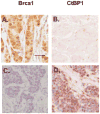
Correlation between CtBP1 up-regulation and Brca1 down-regulation in invasive ductal breast cancers. Immunohistochemical staining was performed using antibodies against Brca1 and CtBP1 to stain consecutive tissue sections as we previously described [20]. Evaluation of CtBP1 and Brca1 staining of human invasive ductal breast cancer samples was performed by 2 independent investigators using methods described previously [20]. Note a lesion with CtBP1 nuclear staining (D) showed negative staining for Brca1 in the consecutive section (C). In contrast, a lesion with CtBP1 staining in the cytoplasm but little in the nucleus (B) exhibited positive Brca1 staining (A). Scale bar = 20μm.
Figure 4
Figure 4
CtBP1 represses E-cadherin expression. A. CtBP1 binding to the E-cadherin regulatory element. MDA-MB-231 cells were used for ChIP assay with an anti-CtBP1 antibody. Primers encompassing the E-cadherin promoter were used to q-PCR-amplify the ChIP sample. * p<0.05 vs. IgG. B. CtBP1 knockdown in MDA-MB-231 cells increased E-cadherin mRNA; * p<0.05 vs. control cells. MDA-MB-231 cells were transfected with scrambled siRNA (Control) or siRNA targeting CtBP1 (siCtBP1) [6] and incubated at 37°C for 48 h. Total RNA was isolated and qRT-PCR was performed as previously described [18]. An 18S probe was used as an internal control. Data are presented as mean ± SEM from a representative experiment. C. CtBP1 knockdown in MDA-MB-231 cells increased E-cadherin protein. Tubulin serves as a loading control. D. CtBP1 knockdown increases E-cadherin expression in MDA-MB-231. Anti E-cadherin was used to stain cells without (SC) or with siCtBP1 treatment (siCtBP1). Scale bar = 15μm.
Figure 5
Figure 5
Correlation between CtBP1 up-regulation and E-cadherin down-regulation in invasive ductal breast cancers. Immunohistochemical staining was performed using antibodies against E-cadherin and CtBP1 to stain consecutive tissue sections as we previously described [20]. Note a lesion without CtBP1 staining (B) exhibited positive E-cadherin staining (A). In contrast, a lesion with CtBP1 nuclear staining (D) showed negative staining for E-cadherin in the consecutive section (C). Scale bar = 40μm.

References

    1. Chinnadurai G. The transcriptional corepressor CtBP: a foe of multiple tumor suppressors. Cancer Res. 2009;69(3):731–734. - PMC - PubMed
    1. Furusawa T, Moribe H, Kondoh H, Higashi Y. Identification of CtBP1 and CtBP2 as corepressors of zinc finger-homeodomain factor deltaEF1. Mol Cell Biol. 1999;19(12):8581–8590. - PMC - PubMed
    1. Grooteclaes M, Deveraux Q, Hildebrand J, Zhang Q, Goodman RH, Frisch SM. C-terminal-binding protein corepresses epithelial and proapoptotic gene expression programs. Proc Natl Acad Sci U S A. 2003;100(8):4568–4573. - PMC - PubMed
    1. Grooteclaes ML, Frisch SM. Evidence for a function of CtBP in epithelial gene regulation and anoikis. Oncogene. 2000;19(33):3823–3828. - PubMed
    1. Mroz EA, Baird AH, Michaud WA, Rocco JW. COOH-terminal binding protein regulates expression of the p16INK4A tumor suppressor and senescence in primary human cells. Cancer Res. 2008;68(15):6049–6053. - PubMed

Publication types