. 2011 Jul 27;133(29):11389-98.
doi: 10.1021/ja204524c. Epub 2011 Jul 5.

Dramatic destabilization of transmembrane helix interactions by features of natural membrane environments

Affiliations

Dramatic destabilization of transmembrane helix interactions by features of natural membrane environments

Heedeok Hong et al. J Am Chem Soc. .

Abstract

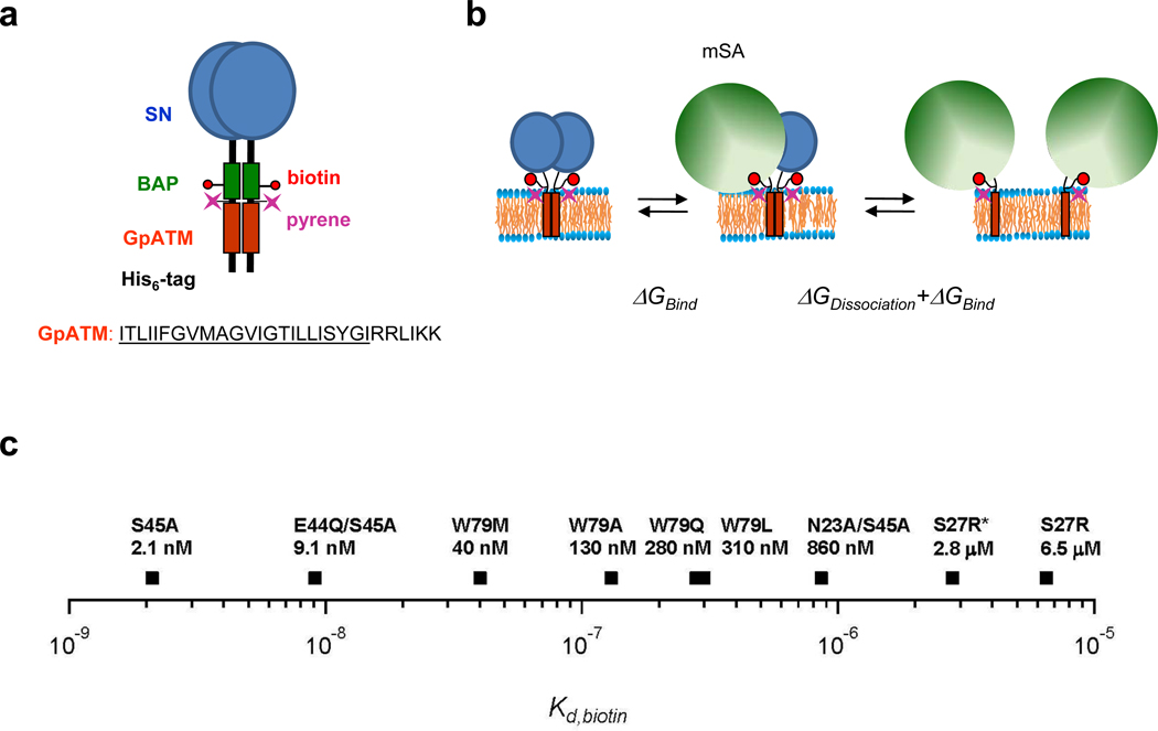
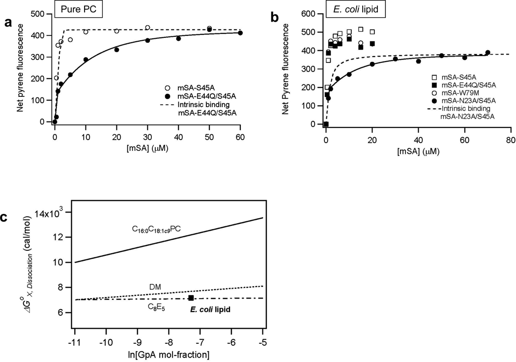
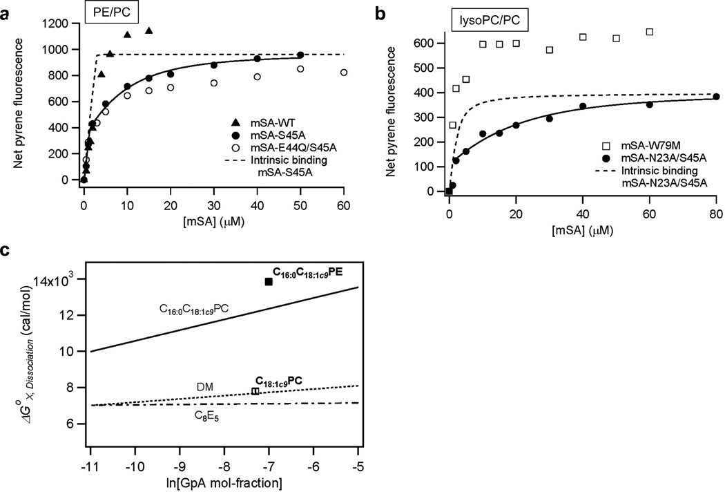
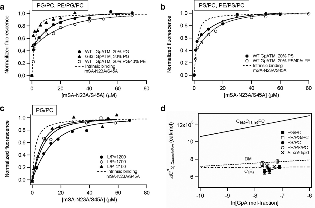
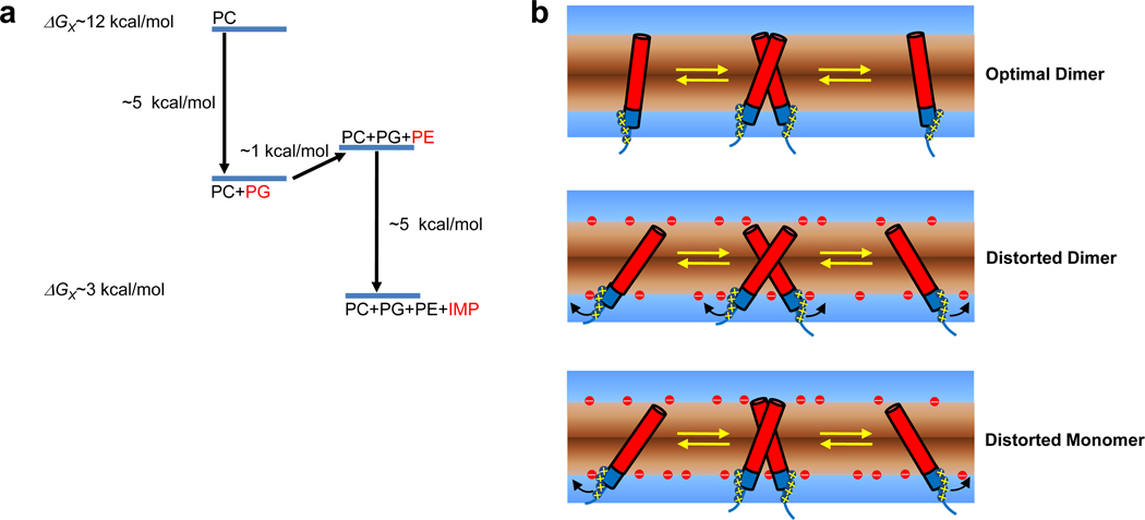
Membrane proteins have evolved to fold and function in a lipid bilayer, so it is generally assumed that their stability should be optimized in a natural membrane environment. Yet optimal stability is not always in accord with optimization of function, so evolutionary pressure, occurring in a complex membrane environment, may favor marginal stability. Here, we find that the transmembrane helix dimer, glycophorin A (GpATM), is actually much less stable in the heterogeneous environment of a natural membrane than it is in model membranes and even common detergents. The primary destabilizing factors are electrostatic interactions between charged lipids and charged GpATM side chains, and nonspecific competition from other membrane proteins. These effects overwhelm stabilizing contributions from lateral packing pressure and excluded volume. Our work illustrates how evolution can employ membrane composition to modulate protein stability.


Figures

Figure 1
Figure 1
The steric trap method. (a) Design of the construct used for steric trapping. GpATM: orange, SN: blue (staphylococcal nuclease fused to the N-terminus), and BAP: green (biotin acceptor peptide). A unique cysteine was labeled with a pyrene fluorescent probe for the detection of binding/dissociation. The amino acid sequence of human GpATM is composed of the hydrophobic transmembrane segment (underlined) followed by a series of positively charged residues in the C-terminus. (b) Reaction scheme of the steric trap method. A single monovalent streptavidin (green sphere) can bind to one of the biotin tags in the dimer with its intrinsic binding affinity (ΔGbind). Because of steric overlap in the dimeric form, however, a second streptavidin can only bind to the dissociated form. Thus, the affinity of the second streptavidin is modulated by the stability of the dimer, yielding an overall free energy of binding equal to ΔGdissociation + ΔGbind. (c) If the affinity of mSA is too high, binding is insensitive to the contribution from dissociation. If the affinity is too low, impractically high concentrations of mSA are required to drive dissociation. We therefore employ a library of mSA mutants with various intrinsic biotin binding affinities (Kd biotin). mSA-S27R (asterisk) has a stronger biotin binding affinity in the negatively charged membranes than in neutral lipid environments (see text).
Figure 2
Figure 2
Comparison of of SNGpA dimer in pure PC and E. coli lipid bilayers. (a) mSA binding curves in pure C16:0C18:1c9PC. The affinity of mSA-S45A is too high under these conditions to observe a second binding phase from dissociation, but the mSA-E44Q/S45A mutant (Kd,biotin=9.1×10⁻⁹ M) allows the dissociation phase to be observed. The fitted Kd,GpA of GpATM dimer was 1.5±0.6×10⁻¹² M at L/P=1000 with mSA-E44Q/S45A. (b) Selection of a mutant mSA with an optimal Kd,biotin to extract the dissociation constant (Kd,GpA) of SNGpATM dimer in E. coli lipid membranes. In contrast to pure PC bilayers, the mSA-E44Q/S45A variant has too high affinity to measure dissociation in E. coli lipid. Instead, a much lower affinity variant was required, mSA-N23A/S45A (Kd,biotin=8.6×10⁻⁷ M), indicative of the weaker dimerization of SNGpATM in E. coli lipids. For the mSA-N23A/S45A binding curve, we obtain Kd,GpA = 1.0±0.3×10⁻⁸ M at a lipid-to-protein molar ratio (L/P) of 1500. All measurements were performed in 20 mM MOPS (pH 7.4), 200 mM NaCl buffer solutions with mSA-accessible SNGpATM concentrations of 1.8 µM. (c) Comparison of the dissociation free energy (ΔGX,Dissociation) of GpATM dimer in E. coli lipid membranes relative to pure C16:0C18:1c9PC bilayers (solid line), DM micelles (dotted line), and C8E5 micelles (dashed line). ΔGX,Dissociation of GpATM dimer is expressed in mol-fraction scale as a function of molar ratio of GpATM to total detergents or lipid concentrations. The lines indicating the concentration dependence of dissociation free energies in various environments were obtained previously,,.
Figure 3
Figure 3
Effects of bilayer lateral pressure profile on the stability of GpATM dimer. (a) mSA-binding curves in C16:0C18:1c9PE/PC (40/60) lipid bilayers. Kd,GpA=2.1±0.6×10⁻¹³ M was obtained at L/P=1100 with mSA-S45A (Kd,biotin=2.1×10⁻⁹ M). (b) mSA binding curves in C18:1c9PC/ C16:0C18:1c9PC (20/80) bilayers. Kd,GpA=6.0±2.4×10⁻⁹ M was obtained at L/P=1500 with mSA-N23A/S45A (Kd,biotin=8.6×10⁻⁷ M). (c) Summary of changes in ΔGX,Dissociation induced by 40 mol-% C16:0C18:1c9PE (filled square) and 20 mol-% C18:1c9PC (empty square) relative to pure C16:0C18:1c9PC bilayers. The lines indicating the concentration dependence of dissociation free energies in various environments were obtained previously,,.
Figure 4
Figure 4
The effect of negatively charged lipids on SNGpATM dimer affinity. (a) mSA binding curves of wild-type and non-dimerizing G83I GpATM mutant in C16:0C18:1c9PG/PC (20/80) and wild-type GpATM in C16:0C18:1c9PE/PG/PC (40/20/40) bilayers at L/P=1200~1300. Kd,GpA=1.4±0.4×10⁻⁸ M and Kd,GpA=5.1±2.2×10⁻⁹ M were obtained in C16:0C18:1c9PG/PC and C16:0C18:1c9PE/PG/PC, respectively, using a weaker biotin binding mSA-N23A/S45A (Kd,biotin=8.6×10⁻⁷M). (b) mSA binding curves of wild-type GpATM in C16:0C18:1c9PS/PC (20/80) and C16:0C18:1c9PE/PS/PC (40/20/40) bilayers at L/P=1800~1900. Kd,GpA=2.5±1.5×10⁻⁸ M and Kd,GpA=1.0±0.4×10⁻⁸ M were obtained in C16:0C18:1c9PS/PC and C16:0C18:1c9PE/PS/PC, respectively, using mSA-N23A/S45A (Kd,biotin=8.6×10⁻⁷M). (c) Dilution effects of GpATM in the negatively charged C16:0C18:1c9PG/PC (20/80) membranes. The fitted Kd,GpA’s were 1.4±0.4×10⁻⁸ M at L/P=1200, 3.2±1.6×10⁻⁸ M at L/P=1700, and 4.7±2.1×10⁻⁸ M at L/P=2100. (d) Summary of the effects of negatively charged lipids. C16:0C18:1c9PG/PC (20/80) and C16:0C18:1c9PS/PC (20/80) destabilize the SNGpATM dimer relative to pure C16:0C18:1c9PC bilayers. A modest stabilization by the introduction of C16:0C18:1c9PE can be seen by the increase of dissociation free energies in C16:0C18:1c9PE/PG/PC (40/20/40) and C16:0C18:1c9PE/PS/PC (40/20/40) bilayers. The stability of GpATM dimer in E. coli lipid membranes is also indicated for comparison. The lines indicating the concentration dependence of dissociation free energies in various environments were obtained previously,,.
Figure 5
Figure 5
Stability of the neutral GpATMRRKK/QQNN in various lipid environments. (a) Binding curves of mSA-S27R to 2 µM wild-type SNGpATM (Kd,GpA=1.7±0.3×10⁻⁷ M) and SNGpATMRRKK/QQNN (Kd,GpA=2.6±0.6×10⁻⁷ M) in 40 mM DM micelles. (b) mSA binding curves of SNGpATMRRKK/QQNN in the negatively charged C16:0C18:1c9PG/PC (20/80) bilayers at L/P=2100. The stability of the dimer was probed using mSA-S45A (Kd,GpA=4.8±2.5×10⁻¹³ M) and mSA-E44Q/S45A (Kd,GpA=7.3±4.1×10⁻¹³ M). (c) mSA binding curves of SNGpATMRRKK/QQNN in neutral C16:0C18:1c9PC bilayers at L/P=1900. The stability of the dimer was probed using mSA-S45A (Kd,biotin=2.1×10⁻⁹ M, Kd,GpA=4.0±2.2×10⁻¹³ M) and mSA-E44Q/S45A (Kd,biotin=9.1×10⁻⁹ M, Kd,GpA=5.8±2.4×10⁻¹³ M).
Figure 6
Figure 6
Effects of total E. coli inner membrane protein extracts (IMP) on the stability of SNGpATM dimer. (a) Binding curves of mSA-S27R to 2.1~2.7 µM SNGpATM with an increasing amount of IMPs in E. coli lipid membranes at L/P=1500. The fitted Kd,GpA’s were 1.1±0.4×10⁻⁸ M and 1.4±0.3×10⁻⁸ M at the IMP to SNGpATM mass ratios of 0 and 0.13, respectively. At higher mass ratios, the dissociation constant was too weak to measure, even with our lowest affinity mSA variant. (b) Changes in the dissociation free energies (ΔGX,Dissociation) of the SNGpATM dimer as a function of the mass ratio of IMPs to SNGpATM in E. coli lipid and C16:0C18:1c9PC membranes. For the detailed experimental design and data, see the Supporting Figure 7.
Figure 7
Figure 7
Summary of the effects of membrane components on GpATM dimer stability. (a) Major environmental forces which contribute to the weak TM helix-helix interactions in natural cell membranes. Relative to neutral fluid C16:0C18:1c9PC bilayers, the negatively charged lipids such as PG and PS dominate the lipid effects, destabilizing the dimer by ~5 kcal/mol. The increased elastic stiffness by the inverted hexagonal phase forming PE lipids mildly stabilizes the dimer. The addition of non-specific proteins to the membranes can further destabilize the specific helix-helix interactions. (b) Possible models for the destabilization of GpATM in the negatively charged membranes. In neutral membrane vesicles, the dimer is stabilized by the restricted lateral and angular motions (top). In the negatively charged membranes, however, interfacial positively charged Arg and Lys residues of TM helices are strongly attracted to the negatively charged lipid headgroups. The resulting adjustment of each monomeric TM helix relative to bilayer normal may cause the distortion of the dimer interface from the optimal side-chain packing destabilizing the dimer (middle). It is also possible that the optimal dimer packing is maintained, but the significant free energy cost is required to form a dimer from a structurally altered, electrostatistically stabilized monomer.

Similar articles

Cited by

References

    1. Bowie JU. Nature. 2005;438:581–589. - PubMed
    1. Andersen OS, Koeppe RE., 2nd Annu Rev Biophys Biomol Struct. 2007;36:107–130. - PubMed
    1. Sachs JN, Engelman DM. Annu Rev Biochem. 2006;75:707–712. - PubMed
    1. Morein S, Andersson A, Rilfors L, Lindblom G. J Biol Chem. 1996;271:6801–6809. - PubMed
    1. Wallin E, von Heijne G. Protein Sci. 1998;7:1029–1038. - PMC - PubMed

Publication types