. 2011 Jul 20;133(28):10748-51.
doi: 10.1021/ja2040656. Epub 2011 Jun 27.

Transpeptidase-mediated incorporation of D-amino acids into bacterial peptidoglycan

Affiliations

Transpeptidase-mediated incorporation of D-amino acids into bacterial peptidoglycan

Tania J Lupoli et al. J Am Chem Soc. .

Abstract

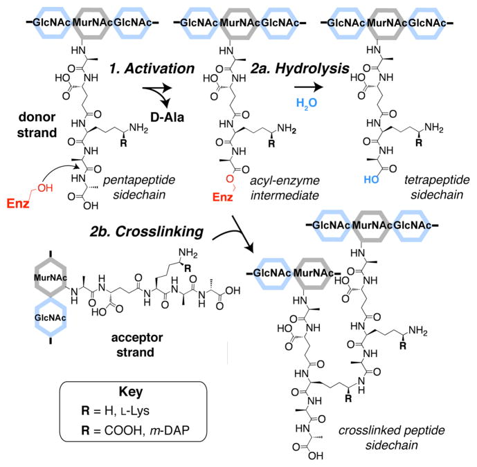
The β-lactams are the most important class of antibiotics in clinical use. Their lethal targets are the transpeptidase domains of penicillin binding proteins (PBPs), which catalyze the cross-linking of bacterial peptidoglycan (PG) during cell wall synthesis. The transpeptidation reaction occurs in two steps, the first being formation of a covalent enzyme intermediate and the second involving attack of an amine on this intermediate. Here we use defined PG substrates to dissect the individual steps catalyzed by a purified E. coli transpeptidase. We demonstrate that this transpeptidase accepts a set of structurally diverse D-amino acid substrates and incorporates them into PG fragments. These results provide new information on donor and acceptor requirements as well as a mechanistic basis for previous observations that noncanonical D-amino acids can be introduced into the bacterial cell wall.


Figures

Figure 1
Figure 1
Stepwise mechanism of peptidoglycan (PG) transpeptidases (TPs). TPs activate PG donor strands (Step 1) resulting in release of terminal D-Ala and formation of an acyl-enzyme intermediate. Either hydrolysis of this activated donor (Step 2a) or peptide crosslinking to an acceptor strand (Step 2b) can then occur. For most Gram-negative bacteria, including E. coli, R=COOH (meso-diaminopimelic acid, m-DAP), while R = H (L-Lys) for most Gram-positive bacteria. (MurNAc: N-acetylmuramic acid; GlcNAc: N-acetylglucosamine)
Figure 2
Figure 2
TPs activate and hydrolyze PG donor strands. (a) Chemical structure of Lipid II analogs and nascent (uncrosslinked) peptidoglycan (PG) where n ~ 30. Amidases cleave peptides from MurNAc residues (dashed red line) of PG. (b) Schematic depicting detection method for modifications to the PG peptide. PG activated by PBP1A* results in release of terminal D-Ala followed by hydrolysis of the acyl-enzyme intermediate, which is indicated by a mixture of tetrapeptides and unreacted pentapeptides upon amidase cleavage. (c) SDS-PAGE analysis showing that E. coli PBP1A* incubated with PG followed by amidase treatment produces pentapeptides and tetrapeptides (lane 4), while PBP1A* inhibited by penicillin G (penG) produces only pentapeptides (lane 6) as seen in the reaction lacking PBP1A* (lane 2). (PBP1A* contains an inactivating mutation, E86Q, in the PGT domain, see SI.)
Figure 3
Figure 3
PG fragments synthesized by E. coli PBP1A are labeled with excess D-Ala via transpeptidation. (a) Top: Chemical reaction depicting transpeptidation of an acyl-enzyme intermediate (donor) and D-amino acid (acceptor) to produce a pentapeptide side chain on PG. Bottom: Schematic of assay to analyze TP product using D-Ala as an acceptor. PG is treated with hydrolases (PBP5 or amidase) followed by paper chromatography to separate unreacted PG (low) from cleaved D-Ala or peptide (high). (c) Assay results: PBP1A (400 nM) was incubated with unlabeled Lipid II (1, 40 μM) and [14C]-D-Ala (1 mM) to produce [14C]-PG. The [14C]-label is removed by treatment with PBP5 or amidase (4 μM each ), but not PBP5 inactivated by penG (5 kU/ml ). Background was not subtracted. Error bars indicate the standard deviation of triplicate experiments.
Figure 4
Figure 4
E. coli PBP1A incorporates D-, but not L-, non-canonical amino acids into the termini of PG peptides via transpeptidation. (a) SDS-PAGE analysis of PBP1A (400 nM) reactions with aromatic amino acids (1 mM) and labeled Ac-Lipid II (2, 4 μM). Amidase treatment of PG products reveals a new peptide band in the D-amino acid reactions (lanes 4, 6, and 8) but not the corresponding L-isomer reactions (lanes 3, 5, and 7) or with added penG (1 kU/ml, lane 1). (b) Overlaid LC-MS extraction traces of amidase-treated PG produced by incubation of D-Phe, D-Tyr, or D-Trp (each 1 mM) with Lipid II (1, 40 μM) and PBP1A (400 nM). Peaks represent mass values of pentapeptides containing aromatic amino acids in place of terminal D-Ala ([M+H]+ calculated, observed m/z values for each, respectively: 565.2980, 565.2984; 581.2930, 581.2933; 604.3089, 604.3085). (c) MS/MS fragmentation of the D-Tyr reaction trace at t = 18.7 min confirms the sequence of the D-Tyr pentapeptide (4) indicated by the chemical structure (inset with calculated m/z values).

References

    1. Scheffers DJ, Pinho MG. Microbiol Mol Biol Rev. 2005;69:585–607. - PMC - PubMed
    2. Sauvage E, Kerff F, Terrak M, Ayala JA, Charlier P. FEMS Microbiol Rev. 2008;32:234–258. - PubMed
    3. Vollmer W, Seligman SJ. Trends Microbiol. 2010;18:59–66. - PubMed
    1. Tipper DJ, Strominger JL. Proc Natl Acad Sci USA. 1965;54:1133–1141. - PMC - PubMed
    2. Ghuysen JM, Frere JM, Leyh-Bouille M, Coyette J, Dusart J, Nguyen-Disteche M. Annu Rev Biochem. 1979;48:73–101. - PubMed
    3. Waxman DJ, Strominger JL. Annu Rev Biochem. 1983;52:825–869. - PubMed
    4. Spratt BG. J Gen Microbiol. 1983;129:1247–1260. - PubMed
    5. Wilke MS, Lovering AL, Strynadka NC. Curr Opin Microbiol. 2005;8:525–533. - PubMed
    6. Testero SA, Fisher JF, Mobashery S. β-Lactam Antibiotics. In Burger’s Medicinal Chemistry. In: Abraham DJ, Rotella DP, editors. Drug Discovery and Development. Vol. 7. Wiley & Sons; New York: 2010. pp. 259–404.
    1. Natural substrates refer to PG (or PG fragments) rather than beta-lactams (see reference 2) or modified peptides (see reference 10). See the following for studies that utilize PG substrates: Waxman DJ, Yu W, Strominger JL. J Biol Chem. 1980;255:11577–11587.Schwartz B, Markwalder JA, Wang Y. J Am Chem Soc. 2001;123:11638–11643.Bertsche U, Breukink E, Kast T, Vollmer W. J Biol Chem. 2005;280:38096–38101.Born P, Breukink E, Vollmer W. J Biol Chem. 2006;281:26985–26993.

    1. The TP domains discussed in this work are fused to N-terminal PGT domains in bifunctional proteins. These TPs had not been shown to catalyze PG hydrolysis using natural substrates. For a review, see: Goffin C, Ghuysen JM. Microbiol Mol Biol Rev. 2002;66:702–738.

    1. For many Gram-positive bacteria, the epsilon-amino group of L-Lys is acylated with other amino acids or peptides, which are involved in cross-links. See the following for a review on PG structure: Vollmer W, Blanot D, de Pedro MA. FEMS Microbiol Rev. 2008;32:149–167.

Publication types

MeSH terms