. 2011 Jun 17:6:41.
doi: 10.1186/1750-1172-6-41.

Incidence, phenotypic features and molecular genetics of Kallmann syndrome in Finland

Affiliations

Incidence, phenotypic features and molecular genetics of Kallmann syndrome in Finland

Eeva-Maria Laitinen et al. Orphanet J Rare Dis. .

Abstract

Background: Kallmann syndrome (KS), comprised of congenital hypogonadotropic hypogonadism (HH) and anosmia, is a clinically and genetically heterogeneous disorder. Its exact incidence is currently unknown, and a mutation in one of the identified KS genes has only been found in ~30% of the patients.

Methods: Herein, we investigated epidemiological, clinical, and genetic features of KS in Finland.

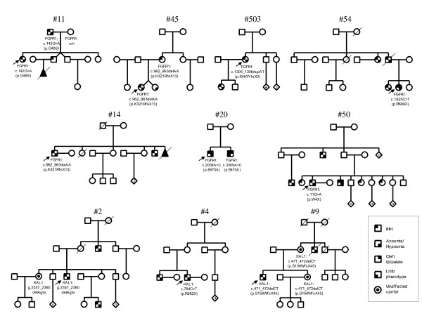
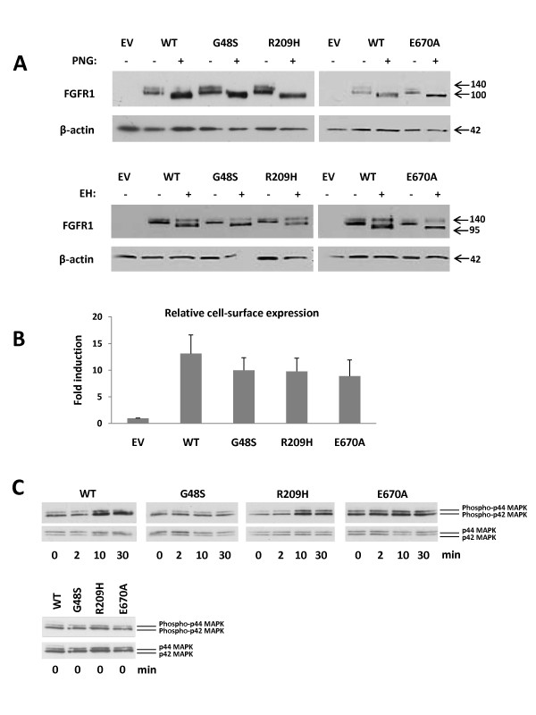
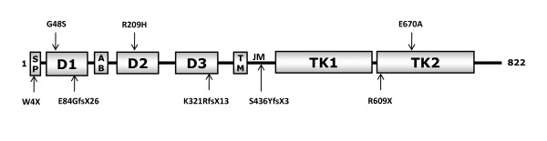
Results: The minimal incidence estimate of KS in Finland was 1:48 000, with clear difference between males (1:30 000) and females (1:125 000) (p = 0.02). The reproductive phenotype of 30 probands (25 men; 5 women) ranged from severe HH to partial puberty. Comprehensive mutation analysis of all 7 known KS genes (KAL1, FGFR1, FGF8, PROK2, PROKR2, CHD7, and WDR11) in these 30 well-phenotyped probands revealed mutations in KAL1 (3 men) and FGFR1 (all 5 women vs. 4/25 men), but not in other genes.

Conclusions: Our results suggest that Finnish KS men harbor mutations in gene(s) yet-to-be discovered with sex-dependent penetrance of the disease phenotype. In addition, some KS patients without CHD7 mutations display CHARGE-syndrome associated phenotypic features (e.g. ear or eye anomalies), possibly implying that, in addition to CHD7, there may be other genes associated with phenotypes ranging from KS to CHARGE.


Figures

Figure 1
Figure 1
Pedigrees of KS patients carrying an FGFR1 or KAL1 mutation. Pedigrees #11, #503, #45, #54, and #50 are consistent with autosomal dominant form of inheritance. X-chromosomal recessive inheritance is apparent in pedigrees #2 and #9.
Figure 2
Figure 2
Functional analyses of FGFR1 mutants. A. Endoglycosidase analysis of mutant FGFR1s. COS-1 cells were transiently transfected with myc-tagged WT or mutated FGFR1 cDNA. EV= empty vector. Cell lysates were subjected to PNGase (PNG, upper panel) or EndoHf (EH, lower panel) digestion. The overall expression of the G48S, R209H and E670A was not significantly decreased as compared to WT (PNGase-treated bands). Receptor maturation patterns are shown in lower panel. The G48S, R209H and E670A mutants have a similar maturation pattern as WT receptor. B. Cell-surface expression of FGFR1 mutants. COS-1 cells were transiently transfected with myc-tagged WT or mutated FGFR1 cDNA. EV = empty vector. Cell-surface expression levels were determined from fixed cells using an anti-myc primary antibody. Absorbancies were detected at 450 nm. The values on the Y-axis represent fold inductions as compared to the level elicited by EV. The WT, G48S, R209H and E670A have similar cell-surface expression levels. C. MAPK signaling analysis of FGFR1 mutants. L6 myoblasts were transiently transfected with myc-tagged WT or mutated FGFR1 cDNA and treated with FGF2 for 0/2/10/30 min. Phospho-specific antibodies (phospho-p44/42 MAPK) were used to determine phosphorylation of MAPK. To control for equal loading, blots were reprobed using an anti p44/42 MAPK antibody. WT and R209H show clear phosphorylation of MAPK after 10 and 30 minutes of FGF2 treatment. With the mutant receptors G48S and E670A, no clear phosphorylation of MAPK was seen in any of the indicated time points. For comparison of baseline activities, all untreated samples (0 min) were also run on the same gel.
Figure 3
Figure 3
The axial 3D T2-weighted MRI images from the region of the inner ear of (A) a KS patient with normal semicircular canals (asterisk), and (B) a KS patient with hypoplastic semicircular canals (arrow), and an unspecified atrophic area (arrow head) in his retina (C).
Figure 4
Figure 4
Schematic of the FGFR1 mutations at protein level. SP, signal peptide; D1-D3, immunoglobulin-like domains; TM, transmembrane domain; JM, juxtamembrane domain; TK1-2, tyrosine kinase domain (contains two subdomains). The G48S mutation is located in the first immunoglobulin-like domain (D1), involved in the receptor autoinhibition. The R209H mutation is located in D2, responsible for ligand binding and specificity. The E670A mutation lies within TK2, responsible for activating the MAP (mitogen-activated protein) kinase pathway. The nonsense mutations, W4X and R609X, and the frameshift mutations (E84GfsX26, K321RfsX13, S436YfsX3), all lead to premature stop codons.

References

    1. Schwanzel-Fukuda M, Bick D, Pfaff DW. Luteinizing hormone-releasing hormone (LHRH)-expressing cells do not migrate normally in an inherited hypogonadal (Kallmann) syndrome. Brain Res Mol Brain Res. 1989;6:311–326. - PubMed
    1. Cariboni A, Maggi R. Kallmann's syndrome, a neuronal migration defect. Cell Mol Life Sci. 2006;63:2512–2526. doi: 10.1007/s00018-005-5604-3. - DOI - PMC - PubMed
    1. Teixeira L, Guimiot F, Dode C, Fallet-Bianco C, Millar RP, Delezoide AL, Hardelin JP. Defective migration of neuroendocrine GnRH cells in human arrhinencephalic conditions. J Clin Invest. 2010;120:3668–3672. doi: 10.1172/JCI43699. - DOI - PMC - PubMed
    1. Raivio T, Falardeau J, Dwyer A, Quinton R, Hayes FJ, Hughes VA, Cole LW, Pearce SH, Lee H, Boepple P, Crowley WF Jr, Pitteloud N. Reversal of idiopathic hypogonadotropic hypogonadism. N Engl J Med. 2007;357:863–873. doi: 10.1056/NEJMoa066494. - DOI - PubMed
    1. Raivio T, Sidis Y, Plummer L, Chen H, Ma J, Mukherjee A, Jacobson-Dickman E, Quinton R, Van Vliet G, Lavoie H, Hughes VA, Dwyer A, Hayes FJ, Xu S, Sparks S, Kaiser UB, Mohammadi M, Pitteloud N. Impaired fibroblast growth factor receptor 1 signaling as a cause of normosmic idiopathic hypogonadotropic hypogonadism. J Clin Endocrinol Metab. 2009;94:4380–4390. doi: 10.1210/jc.2009-0179. - DOI - PMC - PubMed

Publication types

MeSH terms

Substances

LinkOut - more resources