. 2011 Jun 17:8:46.
doi: 10.1186/1742-4690-8-46.

Blocking premature reverse transcription fails to rescue the HIV-1 nucleocapsid-mutant replication defect

Affiliations

Blocking premature reverse transcription fails to rescue the HIV-1 nucleocapsid-mutant replication defect

James A Thomas et al. Retrovirology. .

Abstract

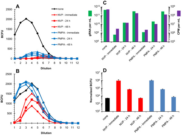
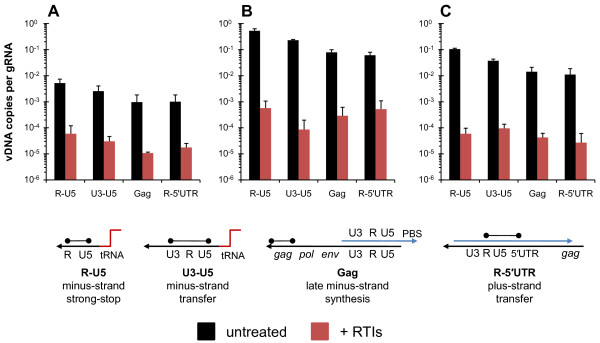
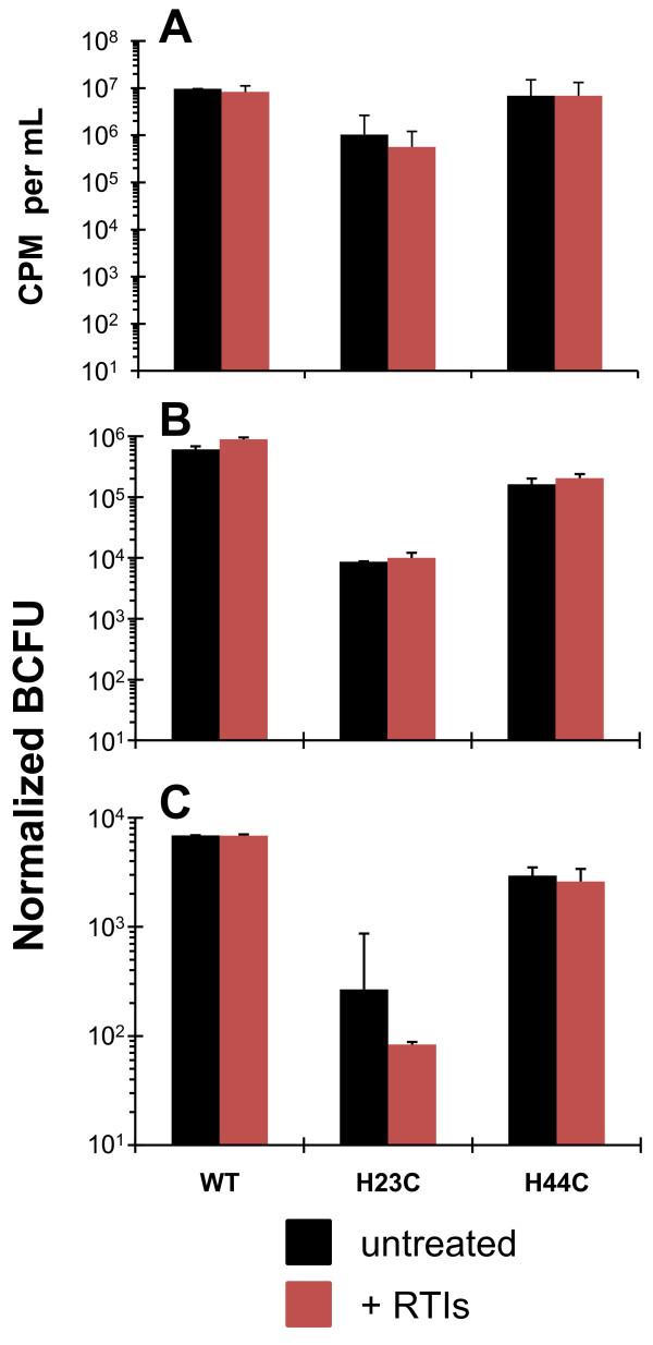
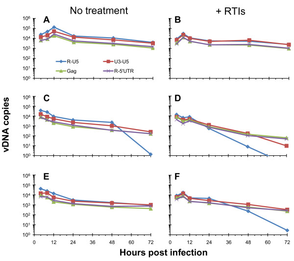
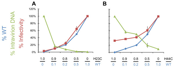
Background: The nucleocapsid (NC) protein of HIV-1 is critical for viral replication. Mutational analyses have demonstrated its involvement in viral assembly, genome packaging, budding, maturation, reverse transcription, and integration. We previously reported that two conservative NC mutations, His23Cys and His44Cys, cause premature reverse transcription such that mutant virions contain approximately 1,000-fold more DNA than wild-type virus, and are replication defective. In addition, both mutants show a specific defect in integration after infection.

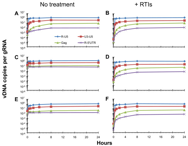
Results: In the present study we investigated whether blocking premature reverse transcription would relieve the infectivity defects, which we successfully performed by transfecting proviral plasmids into cells cultured in the presence of high levels of reverse transcriptase inhibitors. After subsequent removal of the inhibitors, the resulting viruses showed no significant difference in single-round infective titer compared to viruses where premature reverse transcription did occur; there was no rescue of the infectivity defects in the NC mutants upon reverse transcriptase inhibitor treatment. Surprisingly, time-course endogenous reverse transcription assays demonstrated that the kinetics for both the NC mutants were essentially identical to wild-type when premature reverse transcription was blocked. In contrast, after infection of CD4+ HeLa cells, it was observed that while the prevention of premature reverse transcription in the NC mutants resulted in lower quantities of initial reverse transcripts, the kinetics of reverse transcription were not restored to that of untreated wild-type HIV-1.

Conclusions: Premature reverse transcription is not the cause of the replication defect but is an independent side-effect of the NC mutations.


Figures

Figure 1
Figure 1
Methods to remove RTIs from virus preparations. Schematic of the two methods used to remove RTIs from virus preparations. The RTIs were removed so that they did not inhibit downstream assays to assess viral function when premature reverse transcription was blocked. In both methods aspiration was used to remove the supernatant after centrifugation (see the Methods section for details). The method on the left was used to i) maintain competent Env proteins on the surface of virions and ii) limit mechanical stress on virions for subsequent infection analyses. The method on the right uses DNase I treatment to remove extra-virion plasmid DNA contamination with subsequent subtilisin digestion to ensure that the DNase I is completely removed prior to lysis of the virions, and qPCR analysis of intravirion DNA and endogenous reverse transcription assays [33].
Figure 2
Figure 2
RTIs can be effectively removed from virus preparations. Env(-) VSV-G pseudotyped WT HIV-1 expressed from 293T cells transfected in the absence (black), or presence of 1.0 mM PMPA (blue) or 50 μM NVP (red), is assayed for limiting-dilution infectivity on TZM-bl cells (panels A, B and D). The numbers on the X-axes (panels A and B) represent the dilution series, each step being a 3-fold serial dilution starting with 0.1 mL of undiluted infectious supernatant (Dilution 1). For each drug treatment, RTIs were added either immediately before transfection, 24, or 48 h after transfection as indicated in the legend at the right of panel A. Blue colony forming units (BCFU) were tallied 48 h after the infectivity experiments were started [41]. Panel A shows the titer of infectious supernatants assayed without PEG precipitation of virus. Panel B shows the titer of infectious supernatants after PEG precipitation to remove RTIs from the virus stock (see legend from panel A). Panel C shows the yield of WT virus from each transfection condition (with or without drugs) measured using either qRT-PCR (quantifying gRNA per mL, green) or exogenous-template RT assays (measuring RT activity in counts per minute of [32P]-TMP incorporated per mL [CPM per mL], purple). Panel D shows the titer of the PEG-precipitated WT virus, with the treatments (indicated at the bottom) normalized for the gRNA present in the starting supernatant (i.e., corrected for dilution). Values are expressed as normalized blue cell forming units (BCFU) and represent the averages from at least three dilutions (error bars indicate standard deviations).
Figure 3
Figure 3
Premature reverse transcription can be blocked. HIV-1 was expressed from 293T cells transfected either in the absence (black bars) or presence (red bars) of RTIs. Virus was harvested and treated with DNase I and subtilisin, as described in the Methods section (Figure 1, right). Quantities of intravirion DNA were then measured by qPCR using the reverse transcription intermediate targets [31] indicated at the bottom of the figure (tRNA, red line; minus-strand DNA, black line; plus-strand DNA, blue line; target sequences indicated by the black dumbbells). The quantities expressed are the ratio of vDNA to gRNA. Panel A shows wild-type virus, panel B shows NCH23C virus, and panel C shows NCH44C virus. Values plotted are the means and the errors bars are the standard deviations from two separate experiments.
Figure 4
Figure 4
Titer of NC mutant viruses is unchanged if premature reverse transcription is blocked. Results from HIV-1 produced either in the absence (black bars) or the presence (red bars) of RTIs are presented. All viruses produced were PEG precipitated, not just those treated with RTIs for direct comparison. Panel A shows the yields of viruses from transfections, with and without RTI treatment based on average exogenous-template RT activities (in counts per minute of [32P]-TMP incorporated per mL [CPM per mL]; error bars are the standard deviation from duplicate samples). Panels B and C are from two independent transfection-infection experiments and each displays the titers of viruses measured using TZM-bl cells, determined from 3-fold serial dilutions [41]. The titer was corrected for dilution and input virus as determined by exogenous-template RT activity and expressed as "Normalized BCFU" from the means of at least 3 dilutions (error bars represent the standard deviation).
Figure 5
Figure 5
NC mutant viruses display wild-type endogenous reverse transcription kinetics when premature reverse transcription is blocked. Results of endogenous reverse transcription assays performed with virus generated either in the absence or presence of RTIs as indicated at the top of the figure. Virus was prepared using sequential DNase I + subtilisin treatment as described (Figure 1, right). At each time point indicated, a sample of virus was taken and the vDNA was isolated and quantitated. Values were then divided by the number of genomes present at the start of every endogenous reverse transcription reaction to display the quantity of vDNA as a fraction of available genomes. Panels A, C, and E show endogenous reverse transcription time courses using viruses (WT, NCH23C, NCH44C, respectively) prepared without RTIs while panels B, D, and F show time courses with viruses (WT, NCH23C, NCH44C, respectively) prepared in the presence of RTIs. These results are from a representative experiment. The legend for the vDNA species measured is indicated at the bottom of panel A and the bottom of Figure 3 shows schematics of the pertinent vDNA target sites.
Figure 6
Figure 6
NC mutant reverse transcription kinetics in cells are altered when premature reverse transcription is blocked. CD4+ HeLa cells were infected with virus prepared in the absence or presence of RTIs that were subsequently removed using PEG-precipitation (Figure 1, left). These charts display the profile of reverse transcripts over a 72 h time course of infection. Panels A, C, and E show infections from viruses (WT, NCH23C, and NCH44C, respectively) not treated with RTIs, and panels B, D, and F show infections from viruses (WT, NCH23C, and NCH44C, respectively) where premature reverse transcription was blocked via RTI treatment. Prior to the infection, all of the virus samples were normalized for RT activity so that equal amounts were used to infect each set of cells. These results are from a representative experiment. The vDNA species measured were normalized for cell equivalents using CCR5 and are indicated at the bottom of panel A. Schematics of the pertinent vDNA target sites are shown at the bottom of Figure 3.
Figure 7
Figure 7
NCWT phenotype is dominant over NCmutants with no correlation between infectivity and premature reverse transcription. 293T cells were cotransfected with the indicated ratios of NCWT or NCmutant proviral plasmids. The percentage of NCWT in each transfection is shown in blue. The resulting viruses were harvested and infectious titer in TZM-bl cells was determined (red). The resulting titers were corrected for input virus and are expressed as a percentage of WT titer. The amount of intravirion DNA (R-U5) was also quantified in each sample (green), and these values are expressed as a percentage of intravirion DNA present for each respective NCmutant virus (so that 100% mutant is defined as 100% intravirion DNA). The values are the average of 2 separate experiments and the error bars indicate the standard deviations.

Similar articles

Cited by

References

    1. Levin JG, Guo J, Rouzina I, Musier-Forsyth K. Nucleic acid chaperone activity of HIV-1 nucleocapsid protein: Critical role in reverse transcription and molecular mechanism. Prog Nucleic Acid Res Mol Biol. 2005;80:217–286. - PubMed
    1. Rein A, Henderson LE, Levin JG. Nucleic-acid-chaperone activity of retroviral nucleocapsid proteins: significance for viral replication. Trends Biochem Sci. 1998;23:297–301. doi: 10.1016/S0968-0004(98)01256-0. - DOI - PubMed
    1. Thomas JA, Gorelick RJ. Nucleocapsid protein function in early infection processes. Virus Res. 2008;134:39–63. doi: 10.1016/j.virusres.2007.12.006. - DOI - PMC - PubMed
    1. Bampi C, Jacquenet S, Lener D, Decimo D, Darlix JL. The chaperoning and assistance roles of the HIV-1 nucleocapsid protein in proviral DNA synthesis and maintenance. Int J Biochem Cell Biol. 2004;36:1668–1686. doi: 10.1016/j.biocel.2004.02.024. - DOI - PubMed
    1. Bampi C, Jacquenet S, Lener D, Decimo D, Darlix JL. The chaperoning and assistance roles of the HIV-1 nucleocapsid protein in proviral DNA synthesis and maintenance. Curr HIV Res. 2004;2:79–92. doi: 10.2174/1570162043485022. - DOI - PubMed

Publication types

MeSH terms

Substances