

Comparative analysis of macrophage associated vectors for use in genetic vaccine
- PMID: 21682913
- PMCID: PMC3146807
- DOI: 10.1186/1479-0556-9-10
Comparative analysis of macrophage associated vectors for use in genetic vaccine
Abstract
Background: Antigen presentation by non professional antigen presenting cells (APC) can lead to anergy. In genetic vaccines, targeting the macrophages and APC for efficient antigen presentation might lead to balanced immune response. One such approach is to incorporate APC specific promoter in the vector to be used.
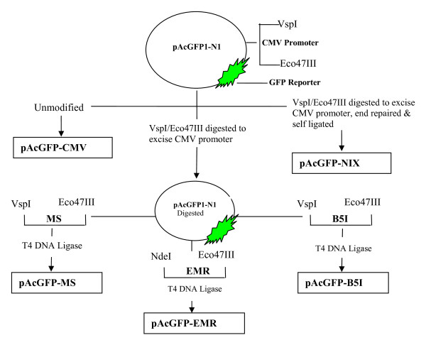
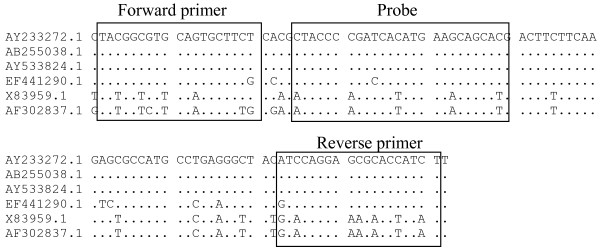
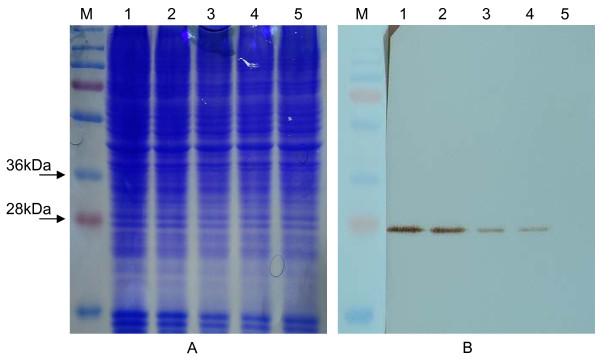
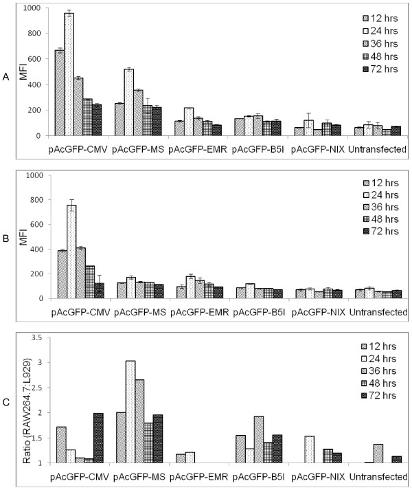
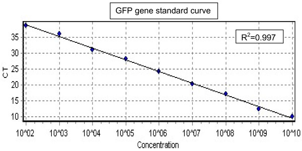
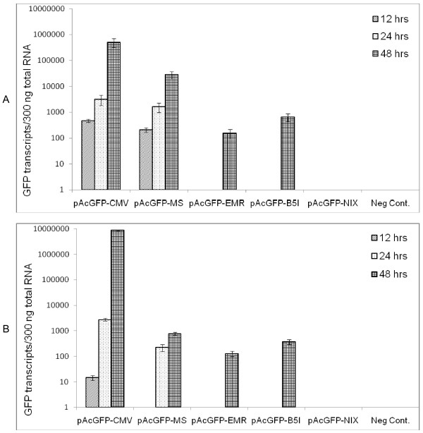
Methods: Three promoters known to be active in macrophage were selected and cloned in mammalian expressing vector (pAcGFP1-N1) to reconstruct (pAcGFP-MS), (pAcGFP-EMR) and (pAcGFP-B5I) with macrosialin, EmrI and Beta-5 Integrin promoters respectively. As a positive control (pAcGFP-CMV) was used with CMV promoter and promoterless vector (pAcGFP-NIX) which served as a negative control. GFP gene was used as readout under the control of each of the promoter. The expression of GFP was analyzed on macrophage and non-macrophage cell lines using Flow cytometry and qRT-PCR with TaqMan probe chemistries.
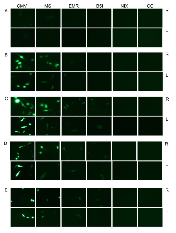
Results: All the promoters in question were dominant to macrophage lineage cell lines as observed by fluorescence, Western blot and quantitative RT-PCR. The activity of macrosialin was significantly higher than other macrophage promoters. CMV promoter showed 1.83 times higher activity in macrophage cell lines. The expression of GFP driven by macrosialin promoter after 24 hours was 4.40 times higher in macrophage derived cell lines in comparison with non macrophage cell lines.
Conclusions: Based on this study, macrosialin promoter can be utilized for targeting macrophage dominant expression. In vivo study needs to be carried out for its utility as a vaccine candidate.
Figures
Similar articles
-
Comparison of immune response generated against Japanese encephalitis virus envelope protein expressed by DNA vaccines under macrophage associated versus ubiquitous expression promoters.Virol J. 2011 Aug 2;8:382. doi: 10.1186/1743-422X-8-382. Virol J. 2011. PMID: 21806845 Free PMC article.
-
[Construction and targeting study of eukaryotic expression vector modulated by a macrophage-specific promoter].Xi Bao Yu Fen Zi Mian Yi Xue Za Zhi. 2008 May;24(5):441-3. Xi Bao Yu Fen Zi Mian Yi Xue Za Zhi. 2008. PMID: 18466696 Chinese.
-
Functional comparison of the murine macrosialin and human CD68 promoters in macrophage and nonmacrophage cell lines.Genomics. 1998 Nov 15;54(1):165-8. doi: 10.1006/geno.1998.5546. Genomics. 1998. PMID: 9806844
-
Lentiviral Vectors Mediate Long-Term and High Efficiency Transgene Expression in HEK 293T cells.Int J Med Sci. 2015 May 15;12(5):407-15. doi: 10.7150/ijms.11270. eCollection 2015. Int J Med Sci. 2015. PMID: 26005375 Free PMC article.
-
Development of a synthetic promoter for macrophage gene therapy.Hum Gene Ther. 2006 Sep;17(9):949-59. doi: 10.1089/hum.2006.17.949. Hum Gene Ther. 2006. PMID: 16972763
Cited by
-
Mannosylated poly(beta-amino esters) for targeted antigen presenting cell immune modulation.Biomaterials. 2015 Jan;37:333-44. doi: 10.1016/j.biomaterials.2014.10.037. Epub 2014 Oct 22. Biomaterials. 2015. PMID: 25453962 Free PMC article.
-
A brief review on DNA vaccines in the era of COVID-19.Future Virol. 2021 Nov:10.2217/fvl-2021-0170. doi: 10.2217/fvl-2021-0170. Epub 2021 Nov 26. Future Virol. 2021. PMID: 34858516 Free PMC article. Review.
-
De novo design of anti-variant COVID-19 vaccine.Biol Methods Protoc. 2023 Sep 26;8(1):bpad021. doi: 10.1093/biomethods/bpad021. eCollection 2023. Biol Methods Protoc. 2023. PMID: 37854896 Free PMC article.
-
Comparison of immune response generated against Japanese encephalitis virus envelope protein expressed by DNA vaccines under macrophage associated versus ubiquitous expression promoters.Virol J. 2011 Aug 2;8:382. doi: 10.1186/1743-422X-8-382. Virol J. 2011. PMID: 21806845 Free PMC article.
-
Engineering of fluorescent or photoactive Trojan probes for detection and eradication of β-Amyloids.Drug Deliv. 2020 Dec;27(1):917-926. doi: 10.1080/10717544.2020.1785048. Drug Deliv. 2020. PMID: 32597244 Free PMC article.
References
-
- Chikhlikar P, Barros de Arruda L, Maciel M, Silvera P, Lewis MG, August JT, Marques ET. DNA encoding an HIV-1 Gag/human lysosome-associated membrane protein-1 chimera elicits a broad cellular and humoral immune response in Rhesus macaques. PLoS One. 2006;1:e135. doi: 10.1371/journal.pone.0000135. - DOI - PMC - PubMed
-
- Li G, Liu Z, Zhong N, Liao B, Xiong Y. Therapeutic effects of DNA vaccine on allergen-induced allergic airway inflammation in mouse model. Cell Mol Immunol. 2006;3(5):379–384. - PubMed
LinkOut - more resources
Full Text Sources
Other Literature Sources
Research Materials
