. 2011 Aug 12;411(2):334-49.
doi: 10.1016/j.jmb.2011.05.050. Epub 2011 Jun 12.

HOXC6 Is transcriptionally regulated via coordination of MLL histone methylase and estrogen receptor in an estrogen environment

Affiliations

HOXC6 Is transcriptionally regulated via coordination of MLL histone methylase and estrogen receptor in an estrogen environment

Khairul I Ansari et al. J Mol Biol. .

Abstract

Homeobox (HOX)-containing gene HOXC6 is a critical player in mammary gland development and milk production, and is overexpressed in breast and prostate cancers. We demonstrated that HOXC6 is transcriptionally regulated by estrogen (E2). HOXC6 promoter contains two putative estrogen response elements (EREs), termed as ERE1(1/2) and ERE2(1/2). Promoter analysis using luciferase-based reporter assay demonstrated that both EREs are responsive to E2, with ERE1(1/2) being more responsive than ERE2(1/2). Estrogen receptors (ERs) ERα and ERβ bind to these EREs in an E2-dependent manner, and antisense-mediated knockdown of ERs suppressed the E2-dependent activation of HOXC6 expression. Similarly, knockdown of histone methylases MLL2 and MLL3 decreased the E2-mediated activation of HOXC6. However, depletion of MLL1 or MLL4 showed no significant effect. MLL2 and MLL3 were bound to the HOXC6 EREs in an E2-dependent manner. In contrast, MLL1 and MLL4 that were bound to the HOXC6 promoter in the absence of E2 decreased upon exposure to E2. MLL2 and MLL3 play key roles in histone H3 lysine-4 trimethylation and in the recruitment of general transcription factors and RNA polymerase II in the HOXC6 promoter during E2-dependent transactivation. Nuclear receptor corepressors N-CoR and SAFB1 were bound in the HOXC6 promoter in the absence of E2, and that binding was decreased upon E2 treatment, indicating their critical roles in suppressing HOXC6 gene expression under nonactivated conditions. Knockdown of either ERα or ERβ abolished E2-dependent recruitment of MLL2 and MLL3 into the HOXC6 promoter, demonstrating key roles of ERs in the recruitment of these mixed lineage leukemias into the HOXC6 promoter. Overall, our studies demonstrated that HOXC6 is an E2-responsive gene, and that histone methylases MLL2 and MLL3, in coordination with ERα and ERβ, transcriptionally regulate HOXC6 in an E2-dependent manner.


Figures

Figure 1
Figure 1
Effect of estrogen on HOXC6 gene expression. (A) JAR cells (grown in phenol red free media) were treated with varying concentrations of E2. RNA from the control and E2-treated cells was isolated, converted to cDNA and analyzed by PCR using primers specific to HOXC6. β-actin was used as a loading control. The cDNA was also analyzed by real-time PCR and expression of HOXC6 (relative to β-actin) is plotted in the right panel. (B) JAR cells were treated with 100 nM E2 for varying time periods (0-24 h) and reverse transcribed-PCR products were analyzed in agarose gel and quantified using real-time PCR (right panel). Each experiment was repeated at least thrice. Bars indicate standard errors (p<0.05).
Figure 2
Figure 2
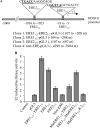
HOXC6 promoter EREs and their estrogen-response: (A) HOX gene promoter EREs (termed as ERE11/2 and ERE21/2, locations and the neighboring sequences are shown). HOXC6 promoter regions spanning ERE11/2 to ERE21/2, ERE11/2 (alone), ERE21/2 (alone) and a non-ERE regions were cloned (clones 1-4) into a luciferase based reporter construct, pGL3, used for transfection and reporter assay. In the mutant pGL3 constructs, clone 1 used for mutation of either ERE11/2 or ERE21/2 alone or both ERE11/2 and ERE21/2 simultaneously. For mutations, the GG of ERE11/2 and CC of ERE21/2 were mutated to AA. (B) Luciferase based reporter assay. ERE11/2-pGL3 and ERE21/2-pGL3 constructs were transfected into JAR cells for 24 h. Control cells were treated with empty pGL3 vector and non-ERE-pGL3. A renilla luciferase construct was also co-transfected along with ERE-pGL3 constructs as an internal transfection control. Cells were then treated with 100 nM E2 and subjected to luciferase assay by using dual-Glo Luciferase Assay System (Promega). The luciferase activities (normalized to renilla activity) in presence of E2 over untreated controls were plotted. The experiment with four replicate treatments was repeated at least twice. Bars indicate standard errors.
Figure 3
Figure 3
Effect of depletion of ERα and ERβ on E2 induced expression of HOXC6. (A) Assessment of ERα and ERβ antisense-mediated knockdown of respective ERs. JAR cells were transfected with ERα, ERβ or scramble antisense (9 μg each) for 48 h and proteins were analyzed by western blot using ERα, ERβ, and β-actin antibodies. (B-C) Effects of ERα and ERβ knockdown on E2-mediated activation of HOXC6. JAR cells were transfected with ERα, ERβ or scramble antisense (9 μg each) for 48 h separately and treated with E2 (100 nM) for additional 8 h. RNA was isolated and subjected to reverse transcriptase-PCR analysis by using primers specific to HOXC6, ERα, and β-actin (loading control). PCR products were analyzed in agarose gel and quantified using real-time PCR (panel C). Lane 1: control cells, lane 2: cells were exposed to 100 nM E2. Lane 3-5: cells were initially transfected with scramble, ERα, and ERβ antisenses separately followed by exposure to E2. Lane 6: Cells were transfected with a mixture (1:1) of ERα and ERβ antisenses followed by exposure to E2. Real-time quantification of cDNA (showing the relative level of HOXC6 expression) is shown in the bottom panel. Each experiment was repeated at least thrice (n = 3). Bars indicate standard errors.
Figure 4
Figure 4
Effect of depletion of MLL1, MLL2, MLL3, and MLL4 on E2-induced expression of HOXC6. JAR cells were transfected with 5 μg (2 × 10⁶ cells) of MLL1, MLL2, MLL3, and MLL4 specific phosphorothioate antisense oligonucleotides separately. Control cells were treated with a phosphorothioate scramble antisense with no homology with MLL1, MLL2, MLL3, and MLL4 genes. The antisense-treated cells were incubated for 48 h followed by treatment with 100 nM E2 for 8 h. RNA was isolated from treated and control cells and subjected to reverse transcriptase-PCR by using primers specific to HOXC6 along with MLL1, MLL2, MLL3, and MLL4. β-actin was used as control. The PCR products were analyzed by agarose gel and quantified. Real-time PCR quantification of the cDNA showing the relative levels of respective MLL and HOXC6 expression are shown in the respective bottom panel. (A) Effects of MLL1 knockdown. (Top) Lane 1: control cells; lane 2: cells that were initially transfected with 5 μg of scramble antisense followed by exposure to E2. Lanes 3: cells were initially transfected with MLL1 antisense and then treated with E2. Expression levels of MLL1 and HOXC6 (relative to actin, average of three replicate experiments, n = 3) were quantified using real-time PCR and plotted in the bottom panel. (B-D) These figures show the effects of knockdown of MLL2, MLL3, and MLL4, respectively, in the similar manner as shown for MLL1 in panel A.
Figure 5
Figure 5
E2-dependent recruitment of ERs and MLLs in the ERE regions of HOXC6 promoter. (A) Scheme showing positions of ChIP PCR primers. (B-C) Recruitment of ERs: JAR cells were treated with 100 nM E2 for 8 h and subjected to ChIP assay using antibodies specific to ERα and ERβ. β-actin antibody was used as control IgG. The immuno-precipitated DNA fragments were PCR-amplified using primers specific to ERE11/2 and ERE21/2 of HOXC6 promoter. Primer specific to a promoter sequence containing no ERE (non-ERE) was used as control. ChIP DNA fragments were analyzed by real-time PCR and shown in the panel B. Each experiment was repeated at least thrice. Bars indicate standard errors. (D-E) Recruitment of MLLs (MLL1-MLL4): JAR cells were treated with 100 nM E2 for 8 h and subjected to ChIP assay using antibodies specific to MLL1, MLL2, MLL3 and MLL4. ChIP DNA fragments were PCR-amplified using primers specific to ERE11/2 and ERE21/2 of HOXC6 promoter. ChIP DNA fragments were analyzed by real-time PCR and shown in panel D. Each experiment was repeated at least thrice. Bars indicate standard errors.
Figure 6
Figure 6
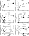
Dynamics of recruitments of ERs and MLLs onto HOXC6 promoter: Cells were treated with 100 nM E2 for varying time periods (0 - 8 h) and then subjected to ChIP assay using antibodies specific to ERα, ERβ, MLL1, MLL2, MLL3, MLL4, H3k4-trimethyl and RNA polymerase II. Immuno-precipitated DNA fragments were PCR-amplified using primers specific to ERE11/2 and ERE21/2 of HOXC6 promoter respectively, quantified and plotted. (A-B) Recruitment of ERα and ERβ in the ERE11/2 and ERE21/2. (C-D) Recruitment of MLL1-4 in the ERE11/2 and ERE21/2. (E-F) Recruitment of RNA pol II (RNAP II) and level of histone H3 (control), H3K4-trimethylation and H3K9-dimethylation. Each experiment was repeated at least thrice. Bars indicate standard errors.
Figure 7
Figure 7
(A-B) Role of MLL2 and MLL3 in E2-dependent assembly of general transcription factors and RNAP II in the HOXC6 promoter. JAR cells were transfected with MLL2, MLL3 or scramble antisenses for 48h and then treated with 100 nM E2 for additional 8h and subjected to ChIP assay by using antibodies specific to H3K4-tri methyl, RNAPII, TBP, TAF250. β-actin antibody was used as control IgG. The immuno-precipitated DNA fragments were PCR-amplified using primers specific to ERE11/2 and ERE21/2 regions of HOXC6 promoter. (C-D) E2-dependent recruitment N-CoR and SAFB1 in ERE regions of HOXC6 promoter in absence and presence of E2. JAR cells were treated with 100 nM E2 for varying time periods (0, 0.5, 4 and 8 h) and subjected to ChIP assay using antibodies specific to N-CoR and SAFB1. Antibodies specific to ERα and β-actin are used as positive and negative control IgG. The ChIP DNA fragments were PCR-amplified using primers specific to ERE11/2 and ERE21/2 of HOXC6 promoter. The real-time PCR quantification of the recruitment level is shown below the respective panels. Each experiment was repeated at least thrice. Bars indicate standard errors.
Figure 8
Figure 8
Roles of ERα and ERβ on E2-dependent recruitment of MLL2 and MLL3. JAR cells were transfected with ERα and ERβ antisense for 48 h followed by exposure to E2 (100 nM for additional 8 h). Cells were harvested and subjected to ChIP assay using anti-MLL2 and anti-MLL3 antibodies. The immuno-precipitated DNA fragments were PCR-amplified using primer specific to ERE11/2 and ERE21/2 regions of HOXC6 promoter and subjected to real-time PCR quantification and plotted (panel B)

Similar articles

Cited by

References

    1. Lappin TR, Grier DG, Thompson A, Halliday HL. HOX genes: seductive science, mysterious mechanisms. Ulster Med J. 2006;75:23–31. - PMC - PubMed
    1. Yu BD, Hess JL, Horning SE, Brown GA, Korsmeyer SJ. Altered Hox expression and segmental identity in Mll-mutant mice. Nature. 1995;378:505–8. - PubMed
    1. Alexander T, Nolte C, Krumlauf R. Hox genes and segmentation of the hindbrain and axial skeleton. Annu Rev Cell Dev Biol. 2009;25:431–56. - PubMed
    1. Chen H, Sukumar S. HOX genes: emerging stars in cancer. Cancer Biol Ther. 2003;2:524–5. - PubMed
    1. Friedmann Y, Daniel CA, Strickland P, Daniel CW. Hox genes in normal and neoplastic mouse mammary gland. Cancer Res. 1994;54:5981–5. - PubMed

Publication types