. 2011 Jul 15;89(1):7-14.
doi: 10.1016/j.ajhg.2011.05.012. Epub 2011 Jun 16.

Mutations in the TGFβ binding-protein-like domain 5 of FBN1 are responsible for acromicric and geleophysic dysplasias

Affiliations

Mutations in the TGFβ binding-protein-like domain 5 of FBN1 are responsible for acromicric and geleophysic dysplasias

Carine Le Goff et al. Am J Hum Genet. .

Abstract

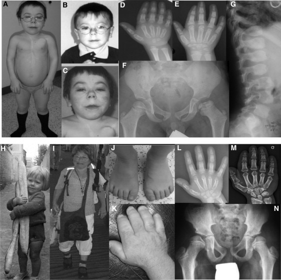
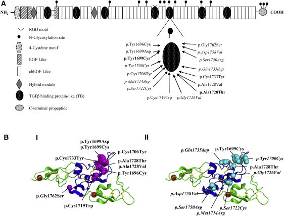
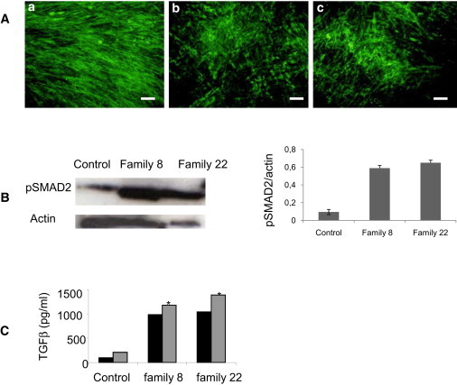
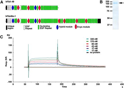
Geleophysic (GD) and acromicric dysplasia (AD) belong to the acromelic dysplasia group and are both characterized by severe short stature, short extremities, and stiff joints. Although AD has an unknown molecular basis, we have previously identified ADAMTSL2 mutations in a subset of GD patients. After exome sequencing in GD and AD cases, we selected fibrillin 1 (FBN1) as a candidate gene, even though mutations in this gene have been described in Marfan syndrome, which is characterized by tall stature and arachnodactyly. We identified 16 heterozygous FBN1 mutations that are all located in exons 41 and 42 and encode TGFβ-binding protein-like domain 5 (TB5) of FBN1 in 29 GD and AD cases. Microfibrillar network disorganization and enhanced TGFβ signaling were consistent features in GD and AD fibroblasts. Importantly, a direct interaction between ADAMTSL2 and FBN1 was demonstrated, suggesting a disruption of this interaction as the underlying mechanism of GD and AD phenotypes. Although enhanced TGFβ signaling caused by FBN1 mutations can trigger either Marfan syndrome or GD and AD, our findings support the fact that TB5 mutations in FBN1 are responsible for short stature phenotypes.


Figures

Figure 1
Figure 1
Clinical and Radiological Features in GD and AD (B, D, F, and G) GD patient 1 at 5 years and (A, C, and E) 15 years; note the “happy” face with full cheeks, a shortened nose, and a long and flat philtrum with a thin upper lip. (D and E) Note the delayed bone age and cone-shaped epiphyses, (F) shortened long tubular bones, epiphyseal dysplasia, and (G) ovoid vertebral bodies. (H and I) AD patient 21 at 3 years and 62 years. Note the round face, bulbous nose, pseudomuscular build, (J, K, L, and M) very short hands and feet with a delayed bone age, and (N) the internal notch of the femoral head.
Figure 2
Figure 2
Location of FBN1 Mutations Identified in GD and AD Patients (A) Functional domains of FBN1. The location of the amino change found in each family is shown (GD are families listed in roman font; AD families are in italics, and mutations shared by AD and GD are in bold). (B) 3D modeling of the fibrillin-1 cbEGF24-TB5-cbEGF25 region showing residues affected by GD and AD mutations. GD substitutions (I) are shown in magenta and AD substitution sites (II) are shown in cyan. Note the clustering of disease-causing substitutions in the region of the TB domain previously associated with protein-protein interactions. cbEGF domains are shown in green, and the TB domain is in blue. Red spheres represent calcium ions bound to domains cbEGF24 and cbEGF25. Homology modeling created with Modeler software and the coordinates of the fibrillin-1 cbEGF22-TB4-cbEGF23 structure (PDB 1UZJ). The figure was generated with Pymol. GD substitutions are shown in roman font, whereas those found in AD or in both diseases are shown in italics and bold, respectively.
Figure 3
Figure 3
Microfibril and TGFβ-Signaling Analysis in GD and AD Skin Fibroblasts (A) Microfibril analysis in skin fibroblasts from (a) control, (b) a GD patient, and (c) an AD patient. The microfibrillar network formation was detected by indirect immunofluorescence with fibrillin-1 antibody. (MAB019). The staining revealed abundant long microfibrils in the control fibroblasts (a). Conversely, the GD and AD patient fibroblasts showed a reduced number of microfibrils and a disorganization of the MF network (b and c). The scale bar represents 50 μm. (B) Enhanced phosphorylation of SMAD2 (pSMAD2) in skin fibroblasts from one GD patient (family 8), one AD patient (family 22), and control. pSMAD2 was normalized to actin for comparison of pSMAD2 levels in affected and unaffected fibroblasts as shown in the right panel. (C) Quantification of total (gray bars) and active (black bars) TGFβ in the conditioned medium of fibroblasts from individuals with GD (family 8) or AD (family 22) and controls. The conditioned medium from families 8 and 22 showed an amount of total TGFβ (p < 0,003) greater than the conditioned medium from the controls.
Figure 4
Figure 4
ADAMTSL2 Interacts Directly with Fibrillin-1 (A) The domain structure of the fibrillin peptide hFib1-49 is shown relative to the full-length fibrillin-1. The key to the fibrillin-1 modules is shown. (B) Colloidal Coomassie-blue-stained-reducing polyacrylamide gel showing purification of hFib1-49 (arrow). The molecular weight markers (in kDa) are indicated on the left. (C) SPR analysis of ADAMTSL2 (analyte) binding to hFib1-49 (ligand). The sensorgrams shown were obtained after injection of increasing concentrations of ADAMTSL2 as indicated. The y axis indicates the response difference obtained between the flow cell with bound hFib1-49 and the control flow cell without hFib1-49 when ADAMTSL2 was used as the analyte. The x axis shows time (s).

References

    1. Spranger J.W., Gilbert E.F., Tuffli G.A., Rossiter F.P., Opitz J.M. Geleophysic dwarfism—a “focal” mucopolysaccharidosis? Lancet. 1971;2:97–98. - PubMed
    1. Maroteaux P., Stanescu R., Stanescu V., Rappaport R. Acromicric dysplasia. Am. J. Med. Genet. 1986;24:447–459. - PubMed
    1. Faivre L., Le Merrer M., Baumann C., Polak M., Chatelain P., Sulmont V., Cousin J., Bost M., Cordier M.P., Zackai E. Acromicric dysplasia: Long term outcome and evidence of autosomal dominant inheritance. J. Med. Genet. 2001;38:745–749. - PMC - PubMed
    1. Le Goff C., Morice-Picard F., Dagoneau N., Wang L.W., Perrot C., Crow Y.J., Bauer F., Flori E., Prost-Squarcioni C., Krakow D. ADAMTSL2 mutations in geleophysic dysplasia demonstrate a role for ADAMTS-like proteins in TGF-beta bioavailability regulation. Nat. Genet. 2008;40:1119–1123. - PMC - PubMed
    1. Allali S., Le Goff C., Pressac-Diebold I., Pfennig G., Mahaut C., Dagoneau N., Alanay Y., Brady A.F., Crow Y.J., Devriendt K. Molecular screening of ADAMTSL2 gene in 33 patients reveals the genetic heterogeneity of geleophysic dysplasia. J. Med. Genet. 2011;48:417–421. - PMC - PubMed

Publication types

MeSH terms

Supplementary concepts