

Benefits of total body photography and digital dermatoscopy ("two-step method of digital follow-up") in the early diagnosis of melanoma in patients at high risk for melanoma
- PMID: 21683472
- PMCID: PMC3215791
- DOI: 10.1016/j.jaad.2011.04.008
Benefits of total body photography and digital dermatoscopy ("two-step method of digital follow-up") in the early diagnosis of melanoma in patients at high risk for melanoma
Abstract
Background: Early detection of melanoma is the best way to improve prognosis. Digital follow-up (DFU) programs of populations at high risk could be an efficient strategy for detecting early melanomas with low morbidity.
Objective: We sought to report the added value of the use of the "two-step method" (digital total body photography and digital dermatoscopy).
Methods: This was an analysis of the surveillance of 618 patients at high risk for melanoma included in our DFU program from 1999 to 2008.
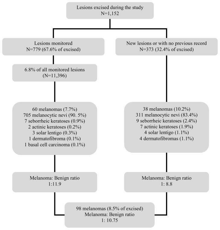
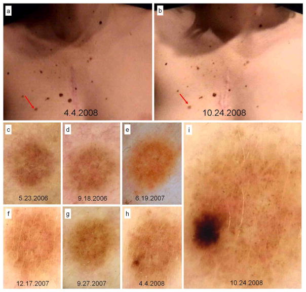
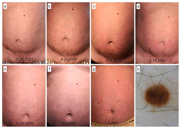
Results: A total of 11,396 lesions were monitored (mean 18.44/patient) during a median follow-up of 96 months (median 10 visits/patient). A total of 1152 lesions, 1.86 per patient, were excised. Almost 70% (798) were lesions previously registered at least twice, whereas 356 (30%) were detected and removed in the same visit. During follow-up, 98 melanomas (8.5% of excised lesions) were diagnosed in 78 patients (12.6%). In all, 53 melanomas were in situ (53.3%), whereas invasive (45) showed a Breslow index of less than 1 mm (median 0.5 mm) and none were ulcerated.
Limitations: Because there are no control groups we cannot determine if the combined use of total body photography and digital dermatoscopy is more beneficial than these techniques used separately.
Conclusion: DFU with total body photography and dermatoscopy in a selected population at high risk demonstrated the early detection of melanomas with a low rate of excisions. Long-term follow-up is required to allow the detection of slow-growing melanomas. Based on our 10-year experience, melanomas can be diagnosed at any time, suggesting that in a population at high risk for melanoma, DFU should be maintained over time.
Copyright © 2011 American Academy of Dermatology, Inc. Published by Mosby, Inc. All rights reserved.
Figures
References
-
- Puig S, Argenziano G, Zalaudek I, et al. Melanomas that failed dermoscopic detection: a combined clinicodermoscopic approach for not missing melanoma. Dermatol Surg. 2007 Oct;33(10):1262–73. - PubMed
-
- Kittler H, Guitera P, Riedl E, et al. Identification of clinically featureless incipient melanoma using sequential dermoscopy imaging. Arch Dermatol. 2006 Sep;142(9):1113–9. - PubMed
-
- Halpern AC. Total body skin imaging as an aid to melanoma detection. Semin Cutan Med Surg. 2003 Mar;22(1):2–8. - PubMed
-
- Malvehy J, Puig S. Follow-up of melanocytic skin lesions with digital total-body photography and digital dermoscopy: a two-step method. Clin Dermatol. 2002 May-Jun;20(3):297–304. - PubMed
Publication types
MeSH terms
Grants and funding
LinkOut - more resources
Full Text Sources
Other Literature Sources
Medical
