. 2011 Jul 22;35(1):34-44.
doi: 10.1016/j.immuni.2011.04.015. Epub 2011 Jun 16.

Anthrax toxin induces macrophage death by p38 MAPK inhibition but leads to inflammasome activation via ATP leakage

Affiliations

Anthrax toxin induces macrophage death by p38 MAPK inhibition but leads to inflammasome activation via ATP leakage

Syed Raza Ali et al. Immunity. .

Abstract

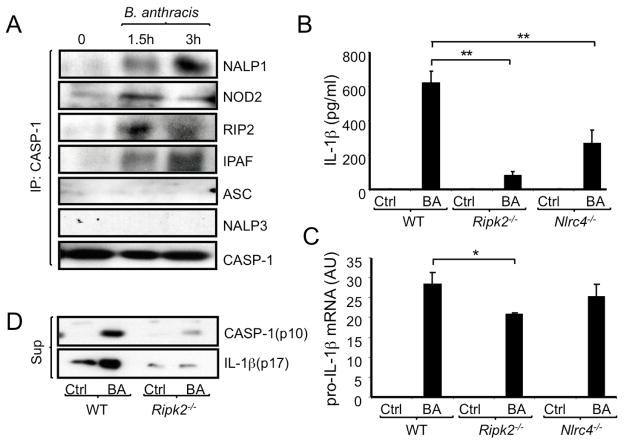
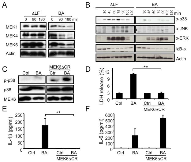
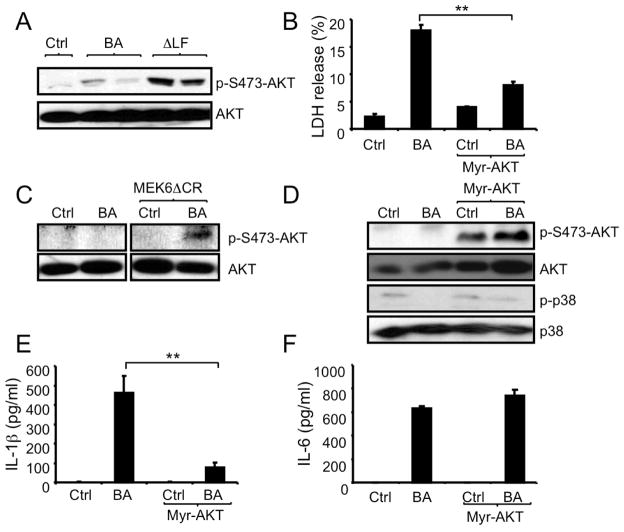
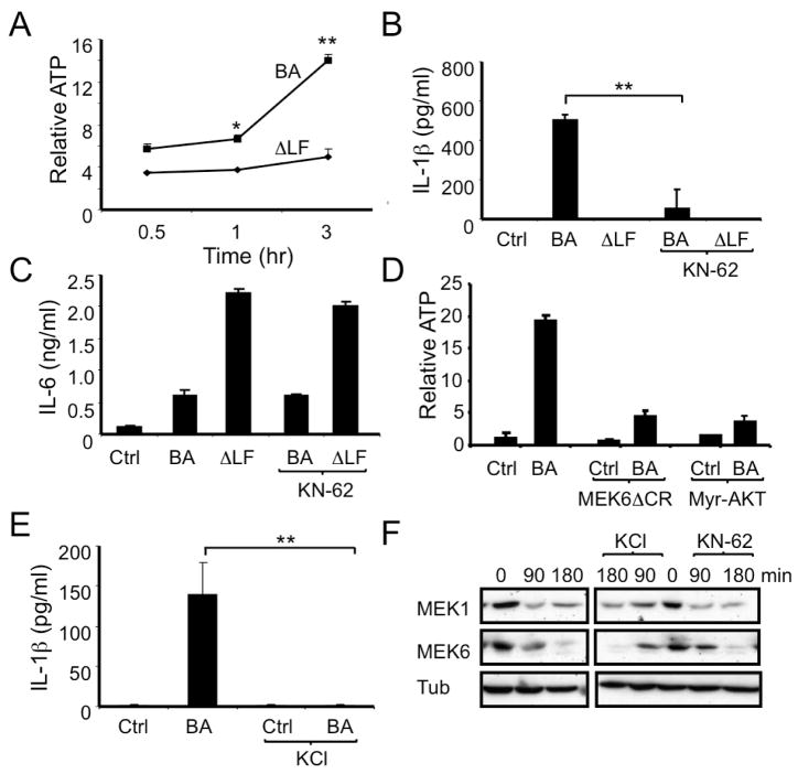
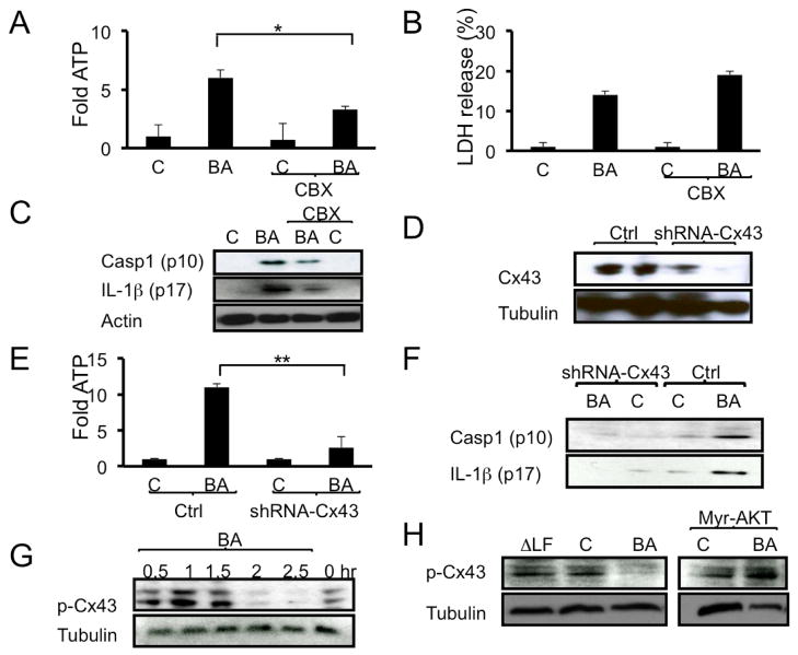
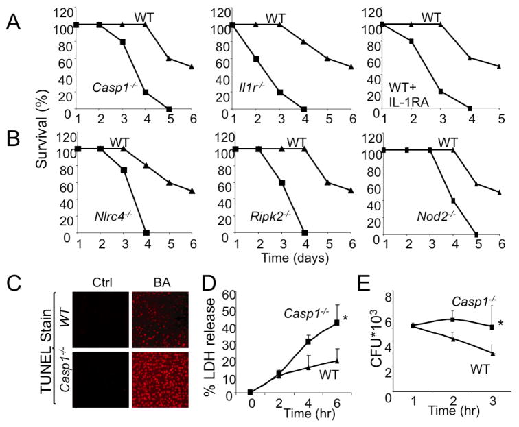
Detection of microbial constituents by membrane associated and cytoplasmic pattern recognition receptors is the essence of innate immunity, leading to activation of protective host responses. However, it is still unclear how immune cells specifically respond to pathogenic bacteria. Using virulent and nonvirulent strains of Bacillus anthracis, we have shown that secretion of ATP by infected macrophages and the sequential activation of the P2X7 purinergic receptor and nucleotide binding oligomerization domain (NOD)-like receptors are critical for IL-1-dependent host protection from virulent B. anthracis. Importantly, lethal toxin produced by virulent B. anthracis blocked activation of protein kinases, p38 MAPK and AKT, resulting in opening of a connexin ATP release channel and induction of macrophage death. Prevention of cell death or ATP release through constitutive p38 or AKT activation interfered with inflammasome activation and IL-1β production, thereby compromising antimicrobial immunity.


Figures

Figure 1
Figure 1. Molecular constituents of B. anthracis-activated inflammasomes
(A) Peritoneal macrophages from C57BL/6 mice were infected with B. anthracis at a multiplicity of infection (MOI) = 2. When indicated, macrophage lysates were prepared and immunoprecipitated (IP) with a caspase-1 antibody. Composition of the immunoprecipitates was examined by immunoblotting of three different blots of the same experiment, which was repeated twice with similar results. (B) Macrophages from the indicated mouse strains were infected with B. anthracis as above. After 8 hr, IL-1β in the culture medium was measured by ELISA. This and all similar experiments were repeated at least 3 times and the results of one representative experiment done in triplicates are shown. Values represent means ± SD. **p < 0.01 denote significant differences between the groups. (C) Macrophages were infected as above and 3 hr later amounts of pro-IL-1β mRNA were measured by quantitative reverse transcriptase polymerase chain reaction (Q-RT-PCR). Results were normalized to the amount of GAPDH mRNA. Values represent means ± SD. *p < 0.05 denotes significant differences between the groups. This experiment was repeated 3 times and the results of one representative experiment done in triplicates are shown. (D) WT and Ripk2−/− macrophages were infected with B. anthracis as above and release of active caspase-1 (p10) and IL-1β (p17) to the culture medium was examined after 12 hr by immunoblotting. This experiment was repeated twice with similar results.
Figure 2
Figure 2. B. anthracis-induced inflammasome activation depends on LF and inhibition of p38
(A,B) C57BL/6 macrophages were infected with WT (BA) or ΔLF B. anthracis. At the indicated times, cell lysates were analyzed by immunoblotting of 2 separate gels of the same experiment for MEK proteolysis (A) and MAPK phosphorylation (B). (C,D) RAW264.7 macrophages were transfected with MEK6ΔCR or an empty vector. Transfected cells were left uninfected or infected with B. anthracis and 1 hr later cell lysates were prepared and analyzed for p38 phosphorylation by immunoblotting (C). Lactate dehydrogenase (LDH) release to the culture supernatant was measured after 4 hr of infection (D). Results are means ± SD, **p < 0.01 denote significant differences between the groups. (E,F) RAW264.7 cells were transfected and infected as above. After 8 hr, IL-1β (E) and IL-6 (F) in culture supernatants were measured by ELISA. Results are means ± SD, **p < 0.01 denote significant differences between the groups. All experiments were repeated at least 3 times and the results of one representative experiment are shown.
Figure 3
Figure 3. B. anthracis-induced cell death and inflammasome activation depend on AKT inhibition
(A) RAW264.7 cells were infected with WT (BA) or ΔLF B. anthracis and 2 hr later, cell lysates were prepared and analyzed by immunoblotting for AKT Ser473 phosphorylation. (B) RAW264.7 cells were retrovirally transduced with myr-AKT or an empty vector. Transduced cells were left uninfected or infected with B. anthracis. LDH in culture supernatants was measured 4 hr later. Results are means ± SD, **p < 0.01 denote significant differences between the groups. (C) RAW264.7 cells were transfected with MEKΔ6CR or an empty vector. Transfected cells were left uninfected or infected with B. anthracis and 2 hr later, cell lysates were prepared and analyzed by immunoblotting for AKT phosphorylation. (D) Myr-AKT-transduced or control RAW264.7 cells were infected with B. anthracis as above. After 1 hr, cell lysates were prepared and analyzed by immunoblotting of 2 separate gels for AKT and p38 phosphorylation. (E,F) RAW264.7 cells transfected with myr-AKT or an empty vector were infected as above. IL-1β (E) and IL-6 (F) in culture supernatants were measured 8 hr after infection by ELISA. Results are means ± SD, **p < 0.01. All experiments were repeated at least 3 times and the results of one representative experiment are shown.
Figure 4
Figure 4. B. anthracis induces ATP release from infected macrophages in LF-, p38- and AKT-dependent manner
(A) C57BL/6 macrophages were infected with WT (BA) or ΔLF B. anthracis. At the indicated times points, ATP in the culture supernatants was measured. This experiment was repeated at least 4 times and the results of one representative experiment done in triplicates are shown. Results are means ± SD, *p < 0.05, **p < 0.01 denote significant differences between the groups. (B,C) Mouse macrophages were infected with WT (BA) or ΔLF B. anthracis in the absence or presence of KN-62 (10 μM). After 8 hr, IL-1β (B) and IL-6 (C) in culture supernatants were measured by ELISA. This experiment was repeated 3 times and the results of one representative experiment done in triplicates are shown. Results are means ± SD, **p < 0.01 denote significant differences between the groups. (D) RAW264.7 cells transfected with MEK6ΔCR, myr-AKT or empty vector were left uninfected or infected with B. anthracis and 2 hr later ATP release to culture supernatants was measured. This experiment was repeated 3 times and the results of one representative experiment done in triplicates are shown. (E) C57BL/6 macrophages were infected with WT B. anthracis in the absence or presence of 130 mM KCl and 8 hr later the amount of IL-1β in culture supernatants was determined by ELISA. This experiment was repeated 3 times and the results of one representative experiment done in duplicates are shown. Results are means ± SD, **p < 0.01 denote significant differences between the groups. (F) Mouse macrophages were infected with WT B. anthracis in the absence or presence of KN-62 (10 μM) or KCl (130 mM) and at the indicated times cell lysates were prepared and analyzed by immunoblotting of 2 separate gels from the same experiment for MEK proteolysis. This experiment was repeated 3 times and the results of one representative experiment are shown.
Figure 5
Figure 5. B. anthracis-induced ATP release and inflammasome activation depend on connexin-43
(A-C) C57BL/6 macrophages were infected with B. anthracis as above and 45 min later the connexin-43 inhibitor carbenoxolone (CBX, 2.5 μM) was added to the culture medium. ATP release was measured 1 hr later (A) and LDH release was measured after 2 hr (B). Secretion of active caspase-1 (p10) and IL-1β (p17) to the culture medium was examined 12 hr after CBX addition. Cellular actin was used as a control. This experiment was repeated 3 times and the results of one representative experiment are shown. (D) RAW264.7 cells were transduced with lentiviruses expressing connexin-43 shRNA or control shRNA (Ctrl.). Connexin-43 expression in the transduced cells was analyzed by immunoblotting. This experiment was repeated 2 times and the results of one representative experiment are shown. (E,F) RAW264.7 cells were transduced with lentiviruses expressing connexin-43 shRNA or control shRNA. Transduced cells were left uninfected or infected with B. anthracis. ATP release was measured 2 hr later (E). Caspase-1 (p10) and IL-1β (p17) secretion was examined after 12 hr (F). This experiment was repeated twice and the results of one representative experiment are shown. Results are means ± SD, **p < 0.01 denote significant differences between the groups. (G) C57BL/6 macrophages were treated as above and at the indicated times cell lysates were prepared and analyzed by immunoblotting for phosphorylation of connexin-43 at Ser279 and Ser282. This experiment was repeated 3 times and the results of one representative experiment are shown. (H) Myr-AKT-expressing or parental RAW264.7 cells were infected as above. After 2 hr, cell lysates were prepared and analyzed by immunoblotting for connexin-43 phosphorylation and tubulin content. This experiment was repeated 3 times and the results of one representative experiment are shown.
Figure 6
Figure 6. Inflammasome activation protects mice from B. anthracis infection
(A,B) Mice of the indicated genotypes were infected with WT B. anthracis (2×10⁶ cfu/mouse given i.p., n=5–8 mice per group). Where indicated, Anakinra (Kineret, IL-1RA, 100 mg/kg) was s.c. injected every 6 hr over a period of 24 hr post-infection. Mouse survival was analyzed over the course of 6 days post-infection. This experiment was repeated 3 times and the results of one representative experiment are shown. (C) WT and Casp1−/− mice were infected as above and 48 hr later their spleens were collected and examined for cell death by a TUNEL assay. The experiment was repeated twice with similar results. (D) Macrophages from WT and Casp1−/− mice were infected with WT B. anthracis and at the indicated times, LDH release was measured. This experiment was repeated 3 times and the results of one representative experiment done in triplicates are shown. Results are means ± SD, *p < 0.05 denote significant differences between the groups. (E) Macrophages from WT and Casp1−/− mice were infected with WT B. anthracis and at the indicated times intracellular bacterial counts were determined. This experiment was repeated 3 times and the results of one representative experiment done in triplicates are shown. Results are means ± SD, *p < 0.05 denote significant differences between the groups.

References

    1. Boyden ED, Dietrich WF. Nalp1b controls mouse macrophage susceptibility to anthrax lethal toxin. Nat Genet. 2006;38:240–244. - PubMed
    1. Bruey JM, Bruey-Sedano N, Luciano F, Zhai D, Balpai R, Xu C, Kress CL, Bailly-Maitre B, Li X, Osterman A, et al. Bcl-2 and Bcl-XL regulate proinflammatory caspase-1 activation by interaction with NALP1. Cell. 2007;129:45–56. - PubMed
    1. Chekeni FB, Elliott MR, Sandilos JK, Walk SF, Kinchen JM, Lazarowski ER, Armstrong AJ, Penuela S, Laird DW, Salvesen GS, et al. Pannexin 1 channels mediate 'find-me' signal release and membrane permeability during apoptosis. Nature. 2010;467:863–867. - PMC - PubMed
    1. Clarke TC, Williams OJ, Martin PE, Evans WH. ATP release by cardiac myocytes in a simulated ischaemia model: inhibition by a connexin mimetic and enhancement by an antiarrhythmic peptide. Eur J Pharmacol. 2009;605:9–14. - PubMed
    1. Comer JE, Chopra AK, Peterson JW, Konig R. Direct inhibition of T-lymphocyte activation by anthrax toxins in vivo. Infect Immun. 2005;73:8275–8281. - PMC - PubMed

Publication types

MeSH terms