. 2011 Aug 15;356(2):486-95.
doi: 10.1016/j.ydbio.2011.06.002. Epub 2011 Jun 13.

Growth of limb muscle is dependent on skeletal-derived Indian hedgehog

Affiliations

Growth of limb muscle is dependent on skeletal-derived Indian hedgehog

Yvette Bren-Mattison et al. Dev Biol. .

Abstract

During embryogenesis, muscle and bone develop in close temporal and spatial proximity. We show that Indian Hedgehog, a bone-derived signaling molecule, participates in growth of skeletal muscle. In Ihh(-/-) embryos, skeletal muscle development appears abnormal at embryonic day 14.5 and at later ages through embryonic day 20.5, dramatic losses of hindlimb muscle occur. To further examine the role of Ihh in myogenesis, we manipulated Ihh expression in the developing chick hindlimb. Reduction of Ihh in chicken embryo hindlimbs reduced skeletal muscle mass similar to that seen in Ihh(-/-) mouse embryos. The reduction in muscle mass appears to be a direct effect of Ihh since ectopic expression of Ihh by RCAS retroviral infection of chicken embryo hindlimbs restores muscle mass. These effects are independent of bone length, and occur when Shh is not expressed, suggesting Ihh acts directly on fetal myoblasts to regulate secondary myogenesis. Loss of muscle mass in Ihh null mouse embryos is accompanied by a dramatic increase in myoblast apoptosis by a loss of p21 protein. Our data suggest that Ihh promotes fetal myoblast survival during their differentiation into secondary myofibers by maintaining p21 protein levels.


Figures

Figure 1
Figure 1. Ptch1 is expressed in muscle tissue and Ihh−/− embryos lose hindlimb skeletal muscle
Wild type and Ihh−/− littermates were harvested at (A) ED14.5, (B) ED16.5, and (C) ED18.5. Hindlimb cross-sections (8μm) were stained with a pan MyHC antibody to detect all differentiated skeletal muscle. F and T mark the fibula and tibia, respectively. (D) Lysates of skeletal muscle tissue from ED16.5 Ihh null and wild type littermates were western blotted for the presence of Ptch1 protein and alpha tubulin was used as a loading control. (E and F) Hindlimb sections were assessed for Ptch1 expression in muscle tissue. (E) Hindlimbs from ED16.5 wild type mice were sectioned and stained with nuclear fast red and X-gal. (F) Hindlimbs from ED16.5 Ptch1+/nLacZ heterozygous littermates were sectioned and stained with nuclear fast red and x-gal to detect beta-galactosidase expression. Inset is enlarged in order to show X-gal+ (blue) muscle tissue( the black spot is an artifact). Dashed lines outline developing muscle tissue (E) and muscle tissue that is β-galactosidase-posistive (F).
Figure 2
Figure 2. Ihh regulates secondary myogenesis
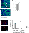
Cross sections at the mid-thigh level of hindlimbs from (A) ED16.5 wild type and (B) Ihh−/− mice stained for MyHC and nuclei reveal larger diameter fibers in Ihh null sections. (A) Arrowheads identify smaller secondary myofibers in sections from wild type limbs. (C) The fiber diameters from sections similar to those shown in (A and B) derived from 3 embryos of each genotype were quantified and the average fiber diameter ± SEM (standard error of the mean) was plotted (*p ≤ 0.01). Myoblasts were explanted from ED20.5 wild type and Ihh−/− hindlimb muscle, plated at equal density, cultured for 3 days and assayed for their ability to form myotubes. (D) Myotubes and nuclei were visualized by using pan MyHC in red and DAPI, respectively. (E) The number of myonuclei per myotube was quantified and plotted as a function of the percent of the total number of myotubes.
Figure 3
Figure 3. Reduction of Ihh expression results in decreased skeletal muscle mass and long bone length
(A) Chick hindlimbs were injected with RCAS(BP)A-Nkx3.2 at ED3 in the mid-region of the hindlimb. (B) On ED12, the limbs were sectioned and an anti-sense Ihh-DIG probe was used to detect Ihh expression following low-dose viral injection. (C and D) Chick hindlimbs were injected with RCAS(BP)A-Nkx3.2 at ED3 and harvested at ED12. (C) Whole-mount immunohistochemistry for MyHC shows decreases in individual muscle sizes and a decrease in girth across the lower limb compared to the contralateral uninfected limb. (D) Extensor muscles (arrows) are not detectable by MyHC staining in Nkx 3.2 infected lower hindlimbs. (E) Forced expression of Nkx3.2 significantly reduces whole limb weight, wet muscle weight and bone length. Chick hindlimbs were injected with RCAS(BP)A-Nkx3.2 on ED6 and harvested on ED12. Both the injected limb and the contralateral control limb were removed and weighed (see experimental procedures). Average percent difference normalized to the contralateral control ± SEM were plotted (*p ≤ 0.01; n=9).
Figure 4
Figure 4. Ectopic Ihh expression increases skeletal muscle mass and rescues Nkx3.2-mediated loss of skeletal muscle mass
(A) Hindlimbs were injected with RCAS(BP)A-Ihh on ED6 and harvested on ED12. Ectopic expression of Ihh increased average whole limb mass and muscle mass without affecting long bone length (for whole limb and muscle mass * is p ≤ 0.01; n=9±SEM). (B) Schematic of co-injection procedure used for rescue of RCAS(BP)A-Nkx3.2 infected limbs with RCAS(BP)A-Ihh. At ED3, both the left and right chick hindlimbs were injected in the mid-region with Nkx3.2 expressing virus. After 24h, an Ihh virus was injected both anteriorly and posteriorly to the site of Nkx3.2 injection in one hindlimb and embryos were maintained until ED10. (C) Whole-mount immunohistochemistry for MyHC of Nkx3.2 and Nkx3.2/Ihh infected hindlimbs. RCAS-Nkx3.2 infection decreased individual muscle size; however, subsequent infection with RCAS-Ihh rescued limb girth and individual muscle sizes (arrows). (D) RCAS-Ihh rescued whole limb and muscle mass loss in Nkx3.2 infected limbs. Average percent difference of Nkx3.2/Ihh co-infected whole limb and wet muscle weight normalized to Nkx3.2 infected contralateral limb were plotted (*p ≤ 0.01; n=5±SEM). (E) Chick hindlimbs were injected with RCAS(BP)A-Nkx3.2 on ED3 and harvested on ED10. Average muscle mass from uninfected contralateral control, RCAS-Nkx3.2 injected, and Nkx3.2/Ihh co-infected limbs (as in D) at ED10 were plotted (uninfected vs. Nkx3.2 single infection; for ED10 harvest whole limb * is p ≤ 0.05; for muscle * is p ≤ 0.01; n=3±SEM).
Figure 5
Figure 5. Ihh-mediated signaling promotes myoblast survival
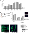
(A) Quantitative PCR analysis was performed on cDNA from wild type littermates and Ihh null hindlimbs harvested on ED16.5 or ED18.5. Muscle regulatory factor (MRF) expression levels were normalized to 18S rRNA and plotted as the average ± SEM. (n ≥3 embryos for each timepoint) (B) Schematic of experiment: wild type chick myoblasts explanted at ED12 were either infected with RCAS(BP)A-Ihh or RCAN control virus. After 24h in culture, cells were switched to differentiation medium and maintained for an additional 72h. Prior to fixation, cells were pulsed with BrdU for 1h and then stained with antibodies to BrdU and MyHC to detect proliferating and differentiated cells, respectively. (C) The data were quantified and the average number of total nuclei, % BrdU+, and % of nuclei in myotubes ± SEMs were plotted per field (30 fields scored per condition; 3 independent experiments; for total nuclei p ≤ 0.05; for % of nuclei in myotubes p ≤ 0.01). Inset micrographs were stained with anti-BrdU in red, anti-MyHC in green and DAPI. (D) Hindlimb cross sections obtained from ED16.5 wild type and Ihh−/− mouse embryos were fixed, and subjected to TUNEL (red) to detect apoptotic cells, and stained for MyHC to detect differentiated muscle (green). Arrows in wild type sections indicate the small number of TUNEL positive cells. (E) Muscle tissue protein extracts were obtained from ED16.5 wild type and Ihh−/− hindlimb muscle, separated by SDS-PAGE and Western blotted. p21 protein was detected in wild type but reduced in Ihh null extracts. Alpha-tubulin was used as a loading control.

Similar articles

Cited by

References

    1. Ahlgren SC, Bronner-Fraser M. Inhibition of sonic hedgehog signaling in vivo results in craniofacial neural crest cell death. Curr Biol. 1999;9:1304–1314. - PubMed
    1. Ayers KL, et al. The long-range activity of Hedgehog is regulated in the apical extracellular space by the glypican Dally and the hydrolase Notum. Dev Cell. 2010;18:605–620. - PubMed
    1. Bader D, et al. Immunochemical analysis of myosin heavy chain during avian myogenesis in vivo and in vitro. J Cell Biol. 1982;95:763–770. - PMC - PubMed
    1. Biressi S, et al. Cellular heterogeneity during vertebrate skeletal muscle development. Dev Biol. 2007;308:281–293. - PubMed
    1. Biressi S, et al. Intrinsic phenotypic diversity of embryonic and fetal myoblasts is revealed by genome-wide gene expression analysis on purified cells. Dev Biol. 2007;304:633–651. - PubMed

Publication types